. 2002 Dec;76(23):11960-70.
doi: 10.1128/jvi.76.23.11960-11970.2002.

Differential regulation of the inhibitor of apoptosis ch-IAP1 by v-rel and the proto-oncogene c-rel

Affiliations

Differential regulation of the inhibitor of apoptosis ch-IAP1 by v-rel and the proto-oncogene c-rel

Jarmila Kralova et al. J Virol. 2002 Dec.

Abstract

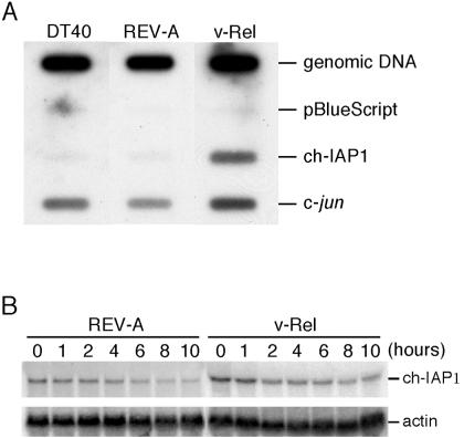
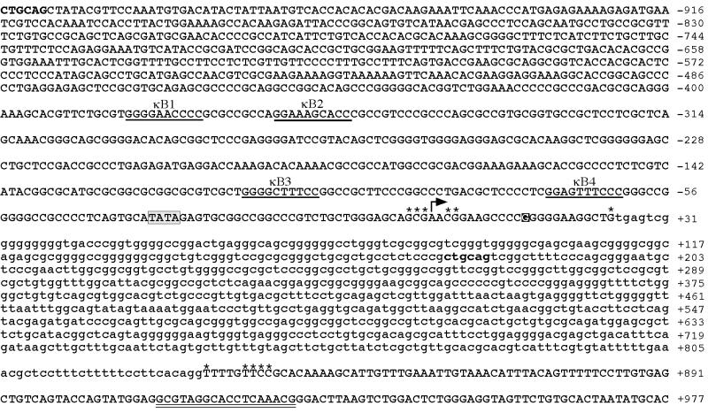
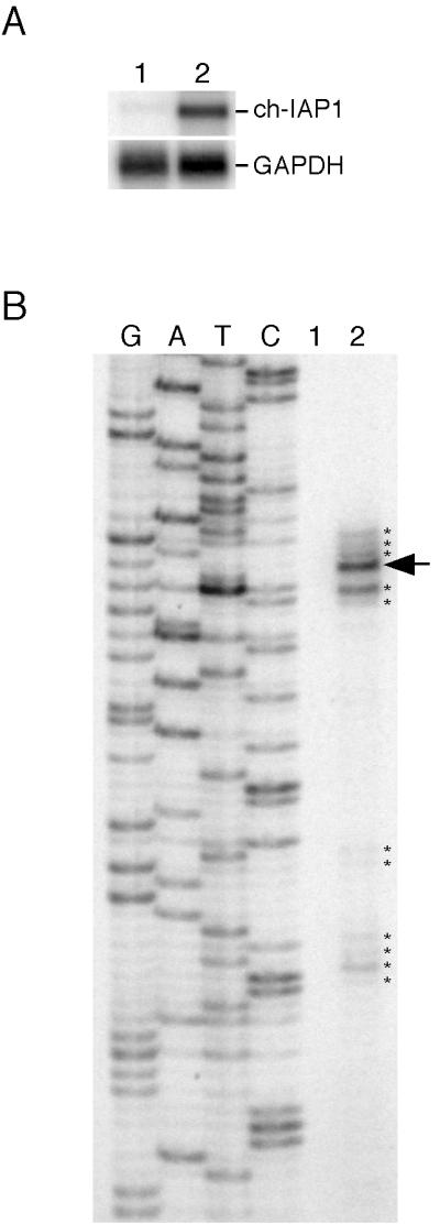
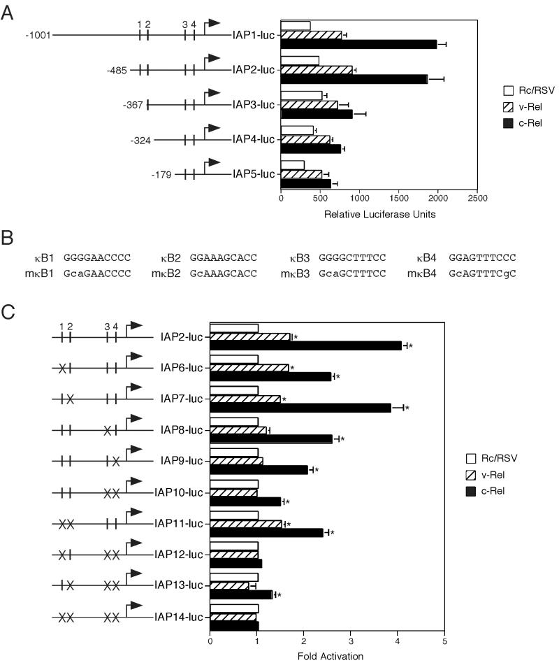
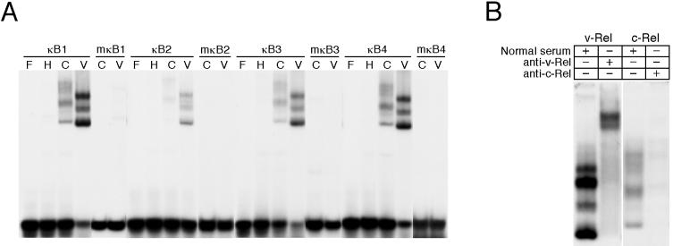
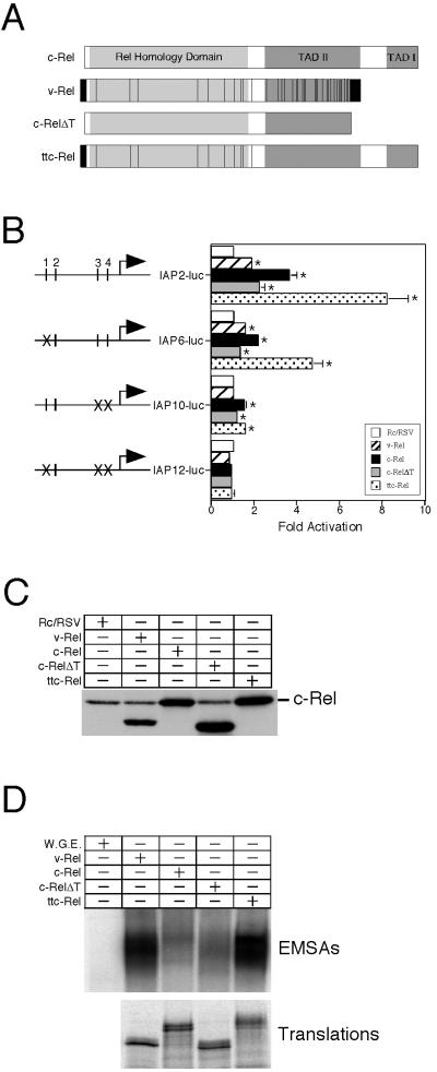
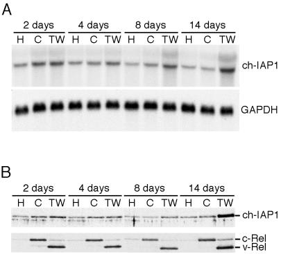
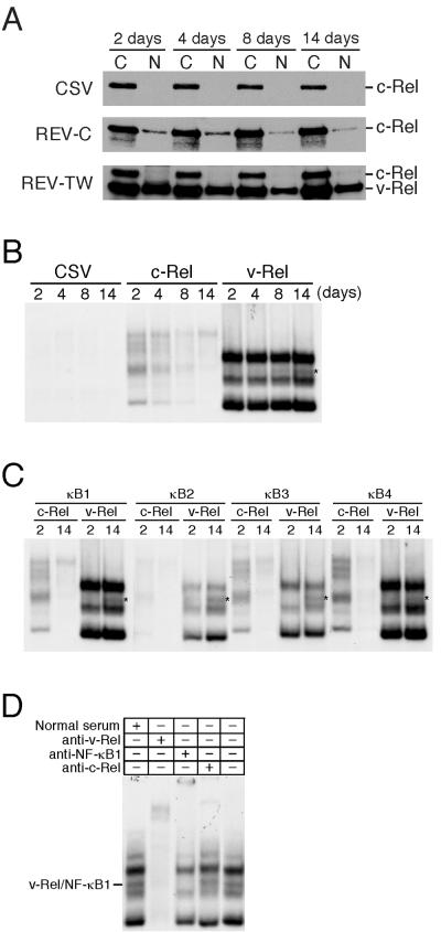
The v-rel oncogene encoded by reticuloendotheliosis virus is the acutely transforming member of the Rel/NF-kappaB family of transcription factors. v-Rel is a truncated and mutated form of c-Rel and transforms cells by inducing the aberrant expression of genes regulated by Rel/NF-kappaB proteins. The expression of ch-IAP1, a member of the inhibitor-of-apoptosis family, is highly elevated in cells expressing v-Rel and contributes to the immortalization of cells transformed by this oncoprotein. In this study we demonstrate that the elevated expression of ch-IAP1 in v-Rel-expressing cells is due to an increased rate of transcription. The ch-IAP1 promoter was isolated, and four Rel/NF-kappaB binding sites were identified upstream of the transcription start site. Two kappaB sites proximal to the transcription start site were required for v-Rel to activate the ch-IAP1 promoter. While c-Rel also utilized these sites, a third more-distal kappaB site was required for its full activation of the ch-IAP1 promoter. Differences in the transactivation domains of v-Rel and c-Rel are responsible for their different abilities to utilize these sites and account for their differential activation of the ch-IAP1 promoter. Although c-Rel was a more potent activator of the ch-IAP1 promoter than v-Rel in transient reporter assays, cells stably overexpressing c-Rel failed to maintain high levels of ch-IAP1 expression. The reduction of ch-IAP1 expression in these cells correlated with the efficient regulation of c-Rel by IkappaBalpha. The ability of v-Rel to escape IkappaBalpha regulation allows for the gradual and sustained elevation of ch-IAP1 expression directly contributing to the transforming properties of v-Rel.


Figures

FIG. 1.
FIG. 1.
Mechanism of increased ch-IAP1 RNA levels in cells expressing v-Rel. DT40 cells were infected with the helper virus REV-A or a retrovirus expressing v-Rel (REV-TW). (A) Nuclear run-on analyses of ch-IAP1 expression. Nuclei isolated from DT40 cells or DT40 cells infected with REV-A or REV-TW were used to produce run-on transcripts in the presence of [32P]UTP. Equivalent amounts of radioactivity from these reactions were hybridized to membranes containing genomic DNA from each cell type, pBluescript plasmid DNA, and plasmids encoding ch-IAP1 and c-jun. Bound radioactivity was visualized by phosphorimager analysis. (B) Half-life analysis of ch-IAP1 RNA in DT40 cells infected with REV-A or REV-TW. Total RNA (10 μg) was isolated from cells treated with actinomycin D (2.5 μg/ml) for 0, 1, 2, 4, 6, 8, and 10 h and analyzed for ch-IAP1 by Northern blot analysis (top panel). Due to low levels of ch-IAP1 mRNA expression in REV-A-infected DT40 cells, a longer exposure of the Northern blot from these cells is shown than for REV-TW-infected cells. ch-IAP1 RNA expression was normalized for loading by comparing the expression of β-actin (lower panel).
FIG. 2.
FIG. 2.
Sequence analysis of the ch-IAP1 promoter. The sequence of a ch-IAP1 genomic clone from −1001 to +977 is shown. The location of the major (arrow) and minor (asterisks) transcription start sites are indicated. The sequence is numbered relative to the major transcription start site (+1). The gray box identifies a potential TATA box at position −33. The nucleotide corresponding to the 5′ end of the ch-IAP1 cDNA is indicated by a black box. Lowercase letters represent sequences encoding the first intron of ch-IAP1, identified by comparison to the ch-IAP1 cDNA. Four potential κB sites (κB1 to -4) are underlined and labeled. Sequences complementary to the primer employed for primer extension are indicated by a double underline. Bold letters indicate the two PstI restriction sites employed to create the IAP1-luc reporter vector.
FIG. 3.
FIG. 3.
Identification of the ch-IAP1 transcription start sites. (A) Expression of ch-IAP1 mRNA in lymphoid cells. Poly(A)+ RNA (1 μg) from MSB-1 (lane 1) or 7.4.1 (lane 2) cells was analyzed for ch-IAP1 expression by Northern blot analysis (top panel). The expression of GAPDH in these cells is shown in the lower panel. (B) Primer extension analysis of the ch-IAP1 mRNA. A probe specific to the ch-IAP1 cDNA was hybridized with poly(A)+ RNA (1 μg) from MSB-1 (lane 1) or 7.4.1 (lane 2) cells and extended with reverse transcriptase. Products of these reactions and DNA sequencing standards were analyzed by denaturing polyacrylamide gel electrophoresis and visualized by phosphorimager analysis. The locations of the major (arrow) and minor (asterisks) primer extension products are indicated.
FIG. 4.
FIG. 4.
v-Rel and c-Rel differentially activate the ch-IAP1 promoter. CEF cultures were transfected with a ch-IAP1 reporter construct, the pRL-TK coreporter vector, and an empty expression plasmid (Rc/RSV) as a control, or Rc/RSV encoding v-Rel or c-Rel. Cells were harvested 32 h after transfection and analyzed for luciferase activity. Luciferase activity from the ch-IAP1 promoter constructs was normalized for transfection efficiency by measuring pRL-TK coreporter activity. The numbers presented are the mean (± standard error) of two to six independent experiments. Reporter activities in extracts from cells overexpressing v-Rel or c-Rel that are significantly different (P < 0.05) from Rc/RSV controls are indicated by an asterisk. (A) Deletion constructs of the ch-IAP1 promoter were subcloned into the pGL3-Basic reporter vector as described in Materials and Methods. Diagrams of the reporter constructs are shown to the left of the luciferase assay results. The relative positions of the κB sites (I) and the major transcription start site (arrow) are indicated. Luciferase assay results are presented as relative luciferase units per microgram of protein. (B) The sequence of the wild-type κB sites (top row) and mutant κB sites (bottom row) are shown. (C) Reporter constructs with wild-type (I) or mutant (×) κB sites were constructed in pGL3-Basic as described in Materials and Methods. The results are presented as fold activation relative to results obtained with an empty expression plasmid control.
FIG. 5.
FIG. 5.
Multiple κB sites in the ch-IAP1 promoter are bound by v-Rel and c-Rel. (A) Nuclear extracts were isolated from CEF cultures 2 days after infection with the helper virus CSV (H), REV-TW (V), or REV-C (C). EMSAs were performed using wild-type or mutant probes encompassing the κB sites found in the ch-IAP1 promoter with 5 μg of nuclear extracts from CSV- or REV-C-infected cells or 1 μg of nuclear extracts from REV-TW-infected cells. The migration profile of free probe (F) for each wild-type κB site is shown. (B) Supershift analysis of κB binding complexes. Nuclear extracts from CEF cultures infected with REV-TW (v-Rel) or REV-C (c-Rel) were incubated with normal rabbit serum or antisera specific for v-Rel or c-Rel prior to the addition of κB3 probe.
FIG. 6.
FIG. 6.
Identification of c-Rel sequences responsible for enhanced activation of the ch-IAP promoter. (A) Diagram of v-Rel and c-Rel proteins. The location of the RHD and transactivation domains I (TAD I) and II (TAD II) are indicated for c-Rel. Vertical lines in v-Rel indicate amino acid differences between v-Rel and c-Rel. The black boxes represent the envelope-derived sequences in v-Rel. A c-Rel construct (c-RelΔT) lacking the 118 C-terminal amino acids missing in v-Rel and a v-Rel construct (ttc-Rel) containing the C-terminal transactivation sequences of c-Rel are shown. (B) Activation of the ch-IAP1 promoter by Rel proteins. CEF cultures were transfected with a ch-IAP1 reporter construct containing wild-type () or mutant (×) κB sites, the pRL-TK coreporter vector, and an empty expression plasmid (Rc/RSV) as a control, or Rc/RSV encoding v-Rel, c-Rel, c-RelΔT, or ttc-Rel. Cells were harvested 32 h after transfectionand analyzed for luciferase activity. Luciferase activity from the ch-IAP1 promoter constructs was normalized for transfection efficiency by measuring pRL-TK coreporter activity. The results presented are the mean fold activation (± standard error) of three independent experiments. Reporter activities in extracts from cells overexpressing Rel proteins that are significantly different (P < 0.05) from Rc/RSV controls are indicated by an asterisk. (C) Expression of transiently expressed Rel proteins. CEF cultures were transfected with an empty Rc/RSV plasmid (1 μg) or Rc/RSV encoding v-Rel, c-Rel, c-RelΔT, or ttc-Rel. Cells lysates were harvested 32 h after transfection and analyzed for the expression of Rel proteins by Western blotting. Exogenously expressed c-Rel and ttc-Rel comigrate with endogenously expressed c-Rel, while v-Rel and c-RelΔT migrate as lower-molecular-weight proteins. (D) DNA binding activity of chimeric Rel proteins. v-Rel, c-Rel, c-RelΔT, or ttc-Rel were translated in vitro, and DNA binding activity was determined by EMSAs using the κB1 site as a probe (top panel). Equal amounts of each protein were used in each reaction as determined by phophorimager analysis of parallel in vitro translation reactions performed with [35S]methionine and were carried out in parallel reactions to show equivalent protein loading in the EMSA analysis (lower panel).
FIG. 7.
FIG. 7.
Kinetic analysis of ch-IAP1 expression. CEF cultures were infected with the helper virus CSV (H), REV-C (C), or REV-TW (TW). Total RNA and whole-cell extracts were isolated at 2, 4, 8, and 14 days after infection. (A) Northern blot analysis of ch-IAP1 expression. Total RNA (10 μg) from cultures infected with CSV, REV-C, or REV-TW was analyzed for the expression of ch-IAP1 RNA (top panel). The expression of GAPDH in these samples is shown to demonstrate the equal loading of RNA (lower panel). (B) Protein from whole-cell lysates (5 × 10⁵ cells) from these cells were resolved by SDS-PAGE, transferred to nitrocellulose, and analyzed by Western blotting with the antiserum specific for ch-IAP1 (top panel) or anti-Rel monoclonal antibody HY87 (lower panel). The migrations of ch-IAP1, v-Rel, and c-Rel are indicated.
FIG. 8.
FIG. 8.
Kinetic analysis of the subcellular localization and κB binding activity of v-Rel and c-Rel. (A) Subcellular localization of v-Rel and c-Rel. Proteins from cytoplasmic (C) and nuclear (N) extracts (5 × 10⁵ cells) from cells infected with CSV (top panel), REV-C (middle panel), or REV-TW (lower panel) were resolved by SDS-PAGE, transferred to nitrocellulose, and analyzed by Western blotting with the anti-Rel monoclonal antibody HY87. The migration of v-Rel and c-Rel are indicated. (B) κB binding activity in cells overexpressing v-Rel or c-Rel. Nuclear extracts were isolated from CEF cultures 2, 4, 8, and 14 days after infection with the helper virus CSV, REV-C (c-Rel), or REV-TW (v-Rel). EMSAs were performed using wild-type probes encompassing the κB1 site found in the ch-IAP1 promoter with 5 μg of nuclear extract from CSV- or REV-C-infected cells or 1 μg of nuclear extract from REV-TW-infected cells. A DNA binding complex that increases in abundance between 2 and 14 days is indicated by an asterisk. (C) EMSAs were performed as described for panel A by usingwild-type probes encompassing the κB1, κB2, κB3, and κB4 sites with nuclear extracts isolated from cells 2 or 14 days after infection. (D) Supershift analysis of κB binding complexes. Nuclear extracts from CEF cultures isolated 14 days after infection with REV-TW were incubated with normal rabbit serum or antisera specific for v-Rel, NF-κB1, or c-Rel prior to the addition of κB1 probe. The binding complex containing v-Rel and NF-κB1 is indicated.

Similar articles

Cited by

References

    1. Ashkenazi, A., and V. M. Dixit. 1998. Death receptors: signaling and modulation. Science 281:1305-1308. - PubMed
    1. Baba, T. W., B. P. Giroir, and E. H. Humphries. 1985. Cell lines derived from avian lymphomas exhibit two distinct phenotypes. Virology 144:139-151. - PubMed
    1. Bose, H. R., Jr. 1992. The Rel family: models for transcriptional regulation and oncogenic transformation. Biochim. Biophys. Acta 1114:1-17. - PubMed
    1. Davis, N., S. Ghosh, D. L. Simmons, P. Tempst, H. C. Liou, D. Baltimore, and H. R. Bose, Jr. 1991. Rel-associated pp40: an inhibitor of the Rel family of transcription factors. Science 253:1268-1271. - PubMed
    1. Diehl, J. A., T. A. McKinsey, and M. Hannink. 1993. Differential pp40IκBβ inhibition of DNA binding by rel proteins. Mol. Cell. Biol. 13:1769-1778. - PMC - PubMed

Publication types

MeSH terms

LinkOut - more resources