. 2002 Dec;76(23):12087-96.
doi: 10.1128/jvi.76.23.12087-12096.2002.

Reassessment of the roles of integrase and the central DNA flap in human immunodeficiency virus type 1 nuclear import

Affiliations

Reassessment of the roles of integrase and the central DNA flap in human immunodeficiency virus type 1 nuclear import

Jeffrey D Dvorin et al. J Virol. 2002 Dec.

Abstract

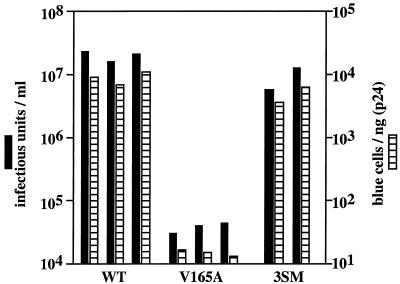
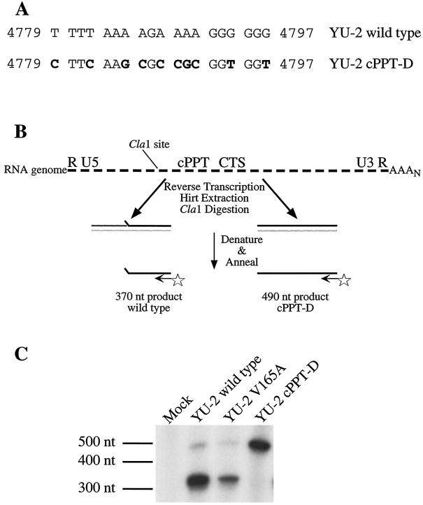
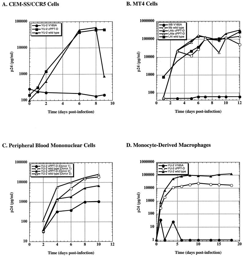
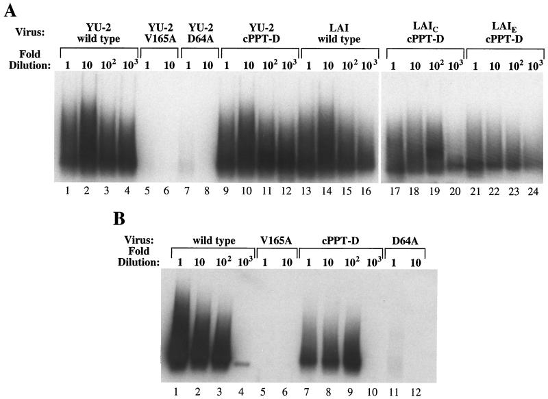
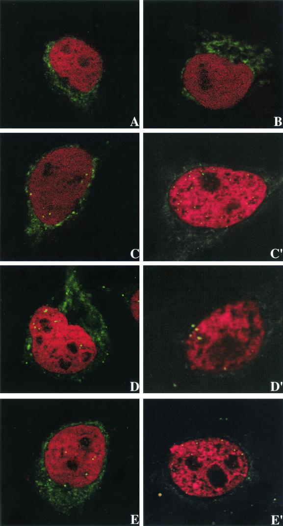
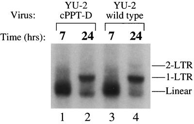
Human immunodeficiency virus type 1 (HIV-1) can infect nondividing cells productively because the nuclear import of viral nucleic acids occurs in the absence of cell division. A number of viral factors that are present in HIV-1 preintegration complexes (PICs) have been assigned functions in nuclear import, including an essential valine at position 165 in integrase (IN-V165) and the central polypurine tract (cPPT). In this article, we report a comparison of the replication and infection characteristics of viruses with disruptions in the cPPT and IN-V165. We found that viruses with cPPT mutations still replicated productively in both dividing and nondividing cells, while viruses with a mutation at IN-V165 did not. Direct observation of the subcellular localization of HIV-1 cDNAs by fluorescence in situ hybridization revealed that cDNAs synthesized by both mutant viruses were readily detected in the nucleus. Thus, neither the cPPT nor the valine residue at position 165 of integrase is essential for the nuclear import of HIV-1 PICs.


Figures

FIG. 1.
FIG. 1.
Infectivity of HIV-1 lacking a potential 5′ splice site near IN-V165. Single-cycle infections of either wild-type (WT) virus or viruses carrying the V165A mutation or three silent mutations that eliminate the potential cryptic splice site at V165 (3SM) without changing the amino acid sequence of integrase (see Materials and Methods) were performed. Infectivity was measured with the MAGI assay with viruses harvested after transfection of proviral clones into 293 cells (solid bars) or normalized to p24Gag levels (hatched bars). These data show that the phenotype of V165A is due to a change in the amino acid sequence of the integrase rather than to a change in the cryptic splice site that overlaps the coding region of V165.
FIG. 2.
FIG. 2.
Disruption of cPPT. (A) Nucleotide sequence of the wild-type and mutant cPPTs. Ten nucleotide changes were introduced into the wild-type cPPT (mutations shown in bold) to form the cPPT-D mutant. The 10 mutations resulted in a single amino acid change in the coding region for integrase (lysine at position 188 was replaced with arginine). (B) Diagram of primer extension assay to measure cPPT activity. Following infection, the RNA genome (dashed line) was reverse transcribed to create the viral cDNA. The plus strand of cDNA was synthesized either as two subgenomic, partially overlapping segments (wild-type virus, left side) or as a single full-length molecule (cPPT-D virus, right side). Following restriction with ClaI, the extracted DNAs were denatured in the presence of a 5′-end-labeled, plus-strand-specific primer (arrow with a star at one end). The primer was extended with Taq polymerase until the enzyme reached either the 5′ end of the downstream plus strand (wild-type virus) or the restricted site (cPPT-D virus). CTS, central termination sequence. (C) Polyacrylamide gel electrophoresis of primer extension products. CEM-SS/CCR5 cells (107) were infected with virus (mock, YU-2 wild type, YU-2 V165A, or YU-2 cPPT-D) corresponding to ∼500 ng of p24Gag and incubated for 6 h, and low-molecular-weight DNAs were extracted. Primer extension was performed as in panel B, and the products were denatured and resolved on a native 5% polyacrylamide-Tris-borate-EDTA gel. The major product from YU-2 wild-type and YU-2 V165A (lanes 2 and 3) was 370 nucleotides (nt), whereas the only product from YU-2 cPPT-D (lane 4) was 490 nucleotides.
FIG. 3.
FIG. 3.
Analysis of HIV-1 replication. Cells were challenged with 293T-derived virus stocks, and replication was measured as the accumulation of p24Gag in the supernatant by ELISA. (A) CEM-SS/CCR5 cells were challenged with inocula corresponding to 10 ng of p24Gag of the R5 set of viruses: YU-2 wild type (solid triangles), YU-2 V165A (solid circles), or YU-2 cPPT-D (solid squares). (B) As in panel A, MT-4 cells were challenged with 100 ng of p24Gag of the X4 set of viruses: IIIB wild type (open circles), LAI wild type (solid squares), LAIE cPPT-D (open triangles), LAIC cPPT-D (solid triangles), or IIIB V165A (solid circles). (C) Peripheral blood mononuclear cells from donor 1 and donor 2 were challenged with 1 and 10 ng, respectively, of p24Gag of the R5 viruses: YU-2 wild type [donor 1 (open circles), donor 2 (open triangles)] or YU-2 cPPT-D [donor 1 (solid circles), donor 2 (solid triangles)]. (D) MDMs (in 48-well plates) that had been maintained for 8 days were challenged with 20 ng of p24Gag of the R5 viruses: YU-2 wild type (solid triangles), YU-2 cPPT-D (open circles), or YU-2 V165A (solid circles).
FIG. 4.
FIG. 4.
Analysis of integration efficiency in vivo. (A) Provirus formation by R5 isolates. Cell lysates obtained 15 to 18 h after infection of CEM-SS/CCR5 cells were serially diluted and subjected to nested Alu-PCR. Amplified products were resolved by agarose gel electrophoresis and analyzed by Southern hybridization with an LTR-specific probe, followed by autoradiography. Integrated HIV-1 DNAs were detected as a broad smear (lanes 7 to 11). (B) Provirus formation in MDMs. As in panel A, lysates obtained 18 h after infection of MDMs were subjected to Alu-PCR analysis.
FIG. 5.
FIG. 5.
FISH. HOS-CD4-CCR5 cells were challenged with inocula corresponding to 200 to 400 ng of p24Gag by centrifugal inoculation, incubated for ∼18 h, fixed, treated with RNase, and ethanol dehydrated. The samples were hybridized overnight at 37°C with an HIV-1-specific, biotinylated probe, followed by fluorescein isothiocyanate-avidin and an additional amplification step. The nuclei were counterstained with propidium iodide. The signals were visualized with a Leica confocal laser scanning microscope. Mock-infected cells (A) and cells challenged in the presence of efavirenz (B) served as negative controls. Representative data from two independent experiments are shown for YU-2 wild type (panels C and C′), YU-2 cPPT-D (panels D and D′), and YU-2 V165A (panels E and E′).
FIG. 6.
FIG. 6.
Nuclear accumulation of viral cDNAs: DNA synthesis and nuclear accumulation. Irradiated CEM-SS/CCR5 cells were challenged with wild-type or cPPT-D mutant virus, and low-molecular-weight DNAs were isolated at 7 and 24 h. Low-molecular-weight DNAs were digested with BsgI and subjected to Southern analysis. The bands corresponding to the linear, 1-LTR circle, and 2-LTR circle forms of viral DNA are indicated.

References

    1. Bell, P., L. J. Montaner, and G. G. Maul. 2001. Accumulation and intranuclear distribution of unintegrated human immunodeficiency virus type 1 DNA. J. Virol. 75:7683-7691. - PMC - PubMed
    1. Bouyac-Bertoia, M., J. D. Dvorin, R. A. Fouchier, Y. Jenkins, B. E. Meyer, L. I. Wu, M. Emerman, and M. H. Malim. 2001. HIV-1 infection requires a functional integrase NLS. Mol. Cell 7:1025-1035. - PubMed
    1. Brown, P. O., B. Bowerman, H. E. Varmus, and J. M. Bishop. 1987. Correct integration of retroviral DNA in vitro. Cell 49:347-356. - PubMed
    1. Bukrinskaya, A. G., A. Ghorpade, N. K. Heinzinger, T. E. Smithgall, R. E. Lewis, and M. Stevenson. 1996. Phosphorylation-dependent human immunodeficiency virus type 1 infection and nuclear targeting of viral DNA. Proc. Natl. Acad. Sci. USA 93:367-371. - PMC - PubMed
    1. Bukrinsky, M. I., S. Haggerty, M. P. Dempsey, N. Sharova, A. Adzhubei, L. Spitz, P. Lewis, D. Goldfarb, M. Emerman, and M. Stevenson. 1993. A nuclear localization signal within HIV-1 matrix protein that governs infection of non-dividing cells. Nature 365:666-669. - PMC - PubMed

Publication types

MeSH terms

LinkOut - more resources