. 2002 Dec;76(23):12097-111.
doi: 10.1128/jvi.76.23.12097-12111.2002.

Toward testing the hypothesis that group B coxsackieviruses (CVB) trigger insulin-dependent diabetes: inoculating nonobese diabetic mice with CVB markedly lowers diabetes incidence

Affiliations

Toward testing the hypothesis that group B coxsackieviruses (CVB) trigger insulin-dependent diabetes: inoculating nonobese diabetic mice with CVB markedly lowers diabetes incidence

S Tracy et al. J Virol. 2002 Dec.

Abstract

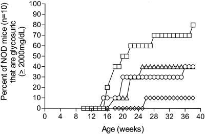
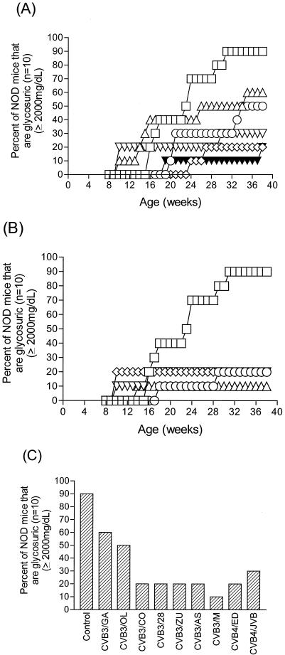
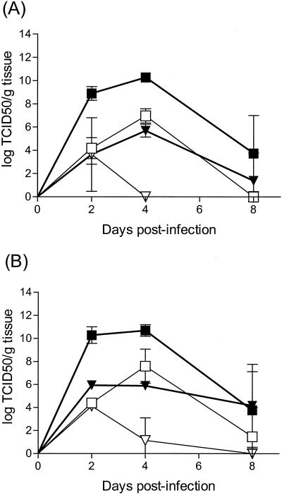
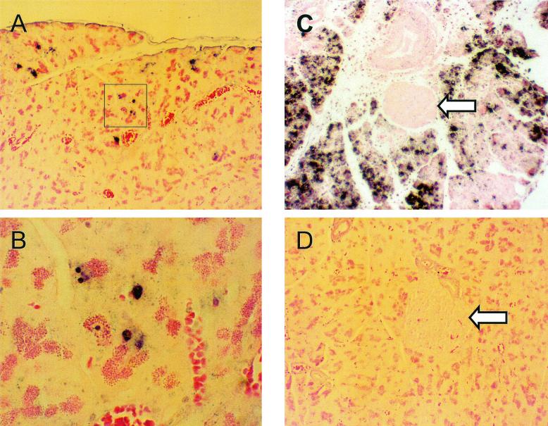
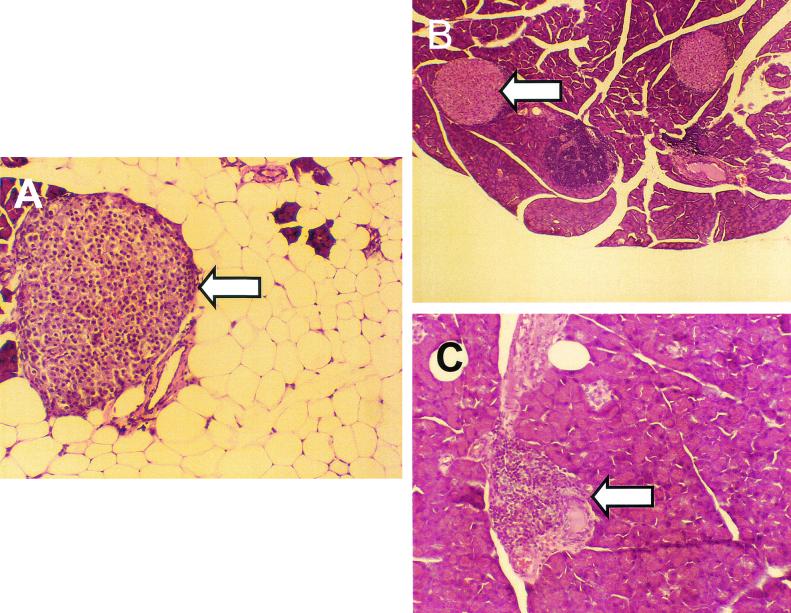
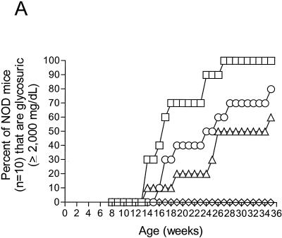
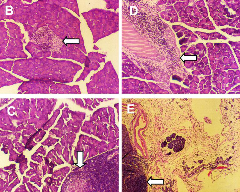
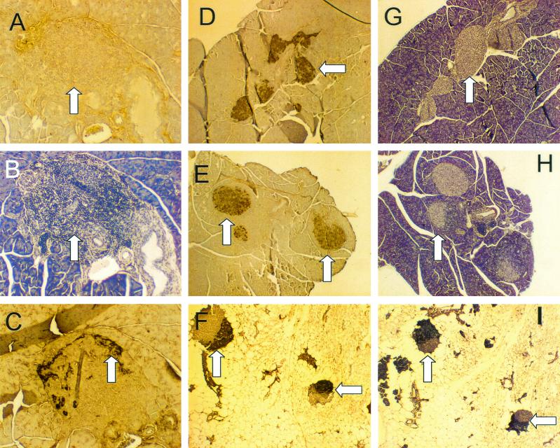
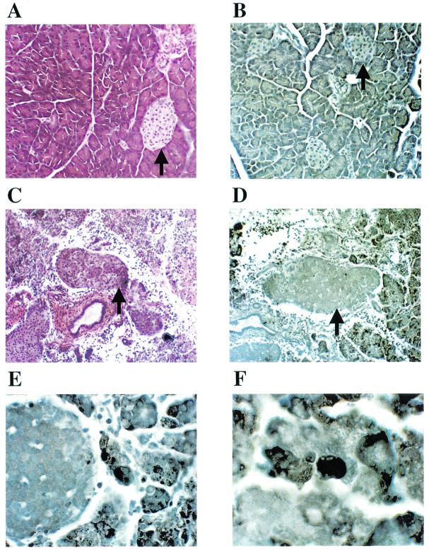
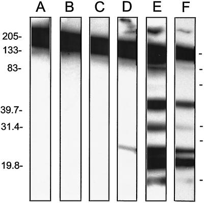
Insulin-dependent (type 1) diabetes mellitus (T1D) onset is mediated by individual human genetics as well as undefined environmental influences such as viral infections. The group B coxsackieviruses (CVB) are commonly named as putative T1D-inducing agents. We studied CVB replication in nonobese diabetic (NOD) mice to assess how infection by diverse CVB strains affected T1D incidence in a model of human T1D. Inoculation of 4- or 8-week-old NOD mice with any of nine different CVB strains significantly reduced the incidence of T1D by 2- to 10-fold over a 10-month period relative to T1D incidences in mock-infected control mice. Greater protection was conferred by more-pathogenic CVB strains relative to less-virulent or avirulent strains. Two CVB3 strains were employed to further explore the relationship of CVB virulence phenotypes to T1D onset and incidence: a pathogenic strain (CVB3/M) and a nonvirulent strain (CVB3/GA). CVB3/M replicated to four- to fivefold-higher titers than CVB3/GA in the pancreas and induced widespread pancreatitis, whereas CVB3/GA induced no pancreatitis. Apoptotic nuclei were detected by TUNEL (terminal deoxynucleotidyltransferase-mediated dUTP-biotin nick end labeling) assay in CVB3/M-infected pancreata but not in CVB3/GA-infected pancreata. In situ hybridization detected CVB3 RNA in acinar tissue but not in pancreatic islets. Although islets demonstrated inflammatory infiltrates in CVB3-protected mice, insulin remained detectable by immunohistochemistry in these islets but not in those from diabetic mice. Enzyme-linked immunosorbent assay-based examination of murine sera for immunoglobulin G1 (IgG1) and IgG2a immunoreactivity against diabetic autoantigens insulin and HSP60 revealed no statistically significant relationship between CVB3-protected mice or diabetic mice and specific autoimmunity. However, when pooled sera from CVB3/M-protected mice were used to probe a Western blot of pancreatic proteins, numerous proteins were detected, whereas only one band was detected by sera from CVB3/GA-protected mice. No proteins were detected by sera from diabetic or normal mice. Cumulatively, these data do not support the hypothesis that CVB are causative agents of T1D. To the contrary, CVB infections provide significant protection from T1D onset in NOD mice. Possible mechanisms by which this virus-induced protection may occur are discussed.


Figures

FIG. 1.
FIG. 1.
CVB3 inoculation of young NOD mice suppresses diabetes incidence. Female NOD mice were inoculated intraperitoneally with 5 × 10⁵ TCID50 of the CVB3 strains shown at 4 and 6 weeks of age. Control mice received virus diluent only. Suppression of T1D incidence by CVB3 was statistically significant (P = 0.0073). Symbols: □, control; ◊, CVB3/M; ○, CVB3/OL; ▵, CVB3/GA.
FIG. 2.
FIG. 2.
CVB3 and CVB4 inoculation of older, prediabetic NOD mice suppresses diabetes incidence. Female NOD mice were inoculated intraperitoneally with 5 × 10⁵ TCID50 of the CVB3 and CVB4 strains shown at 8 and 10 weeks of age. (A and B) Kinetics of diabetes onset for three of the less pathogenic CVB3 strains and two CVB4 strains (A) and for the pathogenic CVB3 strains (B). Symbols in panel A: □, control; ▵, CVB3/GA; ○, CVB3/OL; ◊, CVB3/CO; ▾, CVB4/ED; ▿, CVB4/JVB. Symbols in panel B: □, control; ○, B3/28; ◊, B3/ZU; ▿, B3/AS; ▵, B3/M. (C) Bar graph showing the final T1D incidences in each group when the experiment was ended at 10 months of age. Suppression of T1D by the less-pathogenic strains CVB3/OL, CVB3/CO, and CVB3/GA and the two CVB4 strains (in panel A) or by the pathogenic strains CVB3/M, CVB3/20, CVB3/AS, and CVB3/ZU (in panel B) were statistically significant (P = 0.028 and P = 0.000039, respectively).
FIG. 3.
FIG. 3.
Pathogenic CVB3/M replicates to higher titers than does avirulent CVB3/GA in NOD mouse pancreata and heart tissues. Female NOD mice were inoculated once intraperitoneally with 5 × 10⁵ TCID50 CVB3/M or CVB3/GA at 6 weeks of age (A) or 9 weeks of age (B). Symbols: □, GA pancreas; ▪, M pancreas; ▿, GA heart; ▾, M heart. Tissues were excised when mice were sacrificed and homogenized in tissue culture medium, and the titer of replicating virus was determined on HeLa cell monolayers.
FIG. 4.
FIG. 4.
In situ hybridization demonstrates that CVB3/M and CVB3/GA do not replicate productively in pancreatic islets. Female NOD mice were inoculated once with CVB3/M or CVB3/GA and then sacrificed 4 days later. Pancreatic tissue was formalin fixed and paraffin embedded, and 6-μm were sections cut. In situ hybridization was carried out with a negative-strand digoxigenin-labeled riboprobe to detect the positive-stranded CVB3 genome as described in Materials and Methods. (A) CVB/GA replication was detected in discrete and sparse foci (highlighted foci are shown at ×300 magnification in panel B). (C) CVB3/M replicated throughout the acinar tissue but was not observed in islets. (D) A section of a normal uninfected mouse pancreas similarly probed shows no hybridization. All images are shown at ×75 magnification unless otherwise noted. Arrows indicate islets.
FIG. 5.
FIG. 5.
CVB3/M inoculation induced pancreatitis, which resulted in widespread acinar tissue damage and fat replacement. Sections from formalin-fixed and embedded pancreata of mice inoculated at 4 and 6 weeks of age with CVB3/M (A), CVB3/GA (B), or virus diluent only (control; C) and sacrificed at 38 weeks of age were stained with hemotoxylin and eosin. Loss of acinar tissue subsequent to CVB3/M replication is evident in panel A, whereas exocrine tissue appears to be normal in pancreata from mice inoculated with CVB3/GA (panel B compared to panel C). All images captured at ×75 magnification. Arrowheads indicate islets.
FIG. 6.
FIG. 6.
Induction of anti-CVB3 immunity inhibits CVB3/M-induced pancreatitis. (A) Female NOD mice were inoculated intraperitoneally with 5 × 10⁵ TCID50 of CVB3/GA (CVB3/GA) or virus diluent (control) at 4 weeks of age. Symbols: □, control; ○, CVB3/GA-M; ▵, CVB3/GA; ◊, medium-CVB3/M. One group of mice previously inoculated with CVB3/GA was then inoculated with CVB3/M at 6 weeks of age (CVB3/GA-M). A group of age-matched mice that received only virus diluent at 4 weeks of age was inoculated with CVB3/M at 6 weeks (medium-CVB3/M). The experiment was terminated when surviving mice were 35 weeks old. Acinar tissue destruction was not observed in any mouse inoculated with CVB3/GA alone (B) or CVB3/GA followed by CVB3/M (C) or in control mice (D). Fat replacement due to pancreatitis was observed only in mice after inoculation with CVB3/M (E). Images were captured at ×75 magnification. Arrowheads indicate islets. the suppression of T1D incidence by CVB3/M was highly significant (P = 0.0000055), whereas suppression by CVB3/GA or CVB3/GA, followed by that cause by CVB3/M, was not statistically significant (P = 0.064).
FIG. 6.
FIG. 6.
Induction of anti-CVB3 immunity inhibits CVB3/M-induced pancreatitis. (A) Female NOD mice were inoculated intraperitoneally with 5 × 10⁵ TCID50 of CVB3/GA (CVB3/GA) or virus diluent (control) at 4 weeks of age. Symbols: □, control; ○, CVB3/GA-M; ▵, CVB3/GA; ◊, medium-CVB3/M. One group of mice previously inoculated with CVB3/GA was then inoculated with CVB3/M at 6 weeks of age (CVB3/GA-M). A group of age-matched mice that received only virus diluent at 4 weeks of age was inoculated with CVB3/M at 6 weeks (medium-CVB3/M). The experiment was terminated when surviving mice were 35 weeks old. Acinar tissue destruction was not observed in any mouse inoculated with CVB3/GA alone (B) or CVB3/GA followed by CVB3/M (C) or in control mice (D). Fat replacement due to pancreatitis was observed only in mice after inoculation with CVB3/M (E). Images were captured at ×75 magnification. Arrowheads indicate islets. the suppression of T1D incidence by CVB3/M was highly significant (P = 0.0000055), whereas suppression by CVB3/GA or CVB3/GA, followed by that cause by CVB3/M, was not statistically significant (P = 0.064).
FIG. 7.
FIG. 7.
Immunohistochemical analysis of pancreatic tissue for insulin production. Pancreas sections from mice inoculated either with CVB3/GA or CVB3/M and surviving through 10 months of age without T1D were compared to pancreas sections taken from normal (no virus) diabetic mice and young (4-week-old), normal healthy NOD mice for the expression of insulin by immunohistochemical staining. Typical sections were stained for insulin (A and D to F), glucagon (C), or with hemotoxylin to highlight islet inflammatory infiltrates (B and G to I). See the text for discussion. Sections A to C, D and G, E and H, and F and I are serial sections from the same pancreas. Arrowheads indicate islets. Regions staining for insulin in islets are brown (A, D to F); insulin staining occurs in noninflamed islet regions.
FIG. 8.
FIG. 8.
Apoptosis detected by TUNEL staining only in CVB3-infected pancreata. Four-week-old female NOD mice were inoculated with CVB3/GA or CVB3/M. Pancreas sections taken from paraffin-embedded, formalin-fixed pancreata were probed for apoptosis by the TUNEL technique. Pancreata of CVB3/GA infected mice showed no pancreatitis (A) and no apoptotic nuclei (B). Pancreas sections from CVB3/M-infected mice showed active pancreatitis (C) and acinar tissue that stained widely for apoptotic nuclei by TUNEL (D; representative examples are shown at ×300 magnification in E and F). Arrowheads indicate islets.
FIG. 9.
FIG. 9.
Antibodies in sera from CVB3/M-protected NOD mice detect pancreatic proteins. Pancreatic proteins from normal NOD mice were electrophoresed in nonreducing SDS-containing 10 to 15% polyacrylamide gels and then Western blotted. Strips were cut and individually probed with HRP-labeled secondary antibody only (control) (lane A) or pooled sera from normal 4-week-old healthy (nondiabetic) NOD mice (lane B), normal mice that developed diabetes naturally (lane C), CVB3/GA-protected mice (lane D), or CVB3/M-protected mice (E and F). The exposure time for lanes A to E was 10 min; the exposure time for lane F was 3 min. Molecular mass markers are shown on the side in kilodaltons.

References

    1. Akhtar, N., J. Ni, D. Stromberg, G. L. Rosenthal, N. E. Bowles, and J. A. Towbin. 1999. Tracheal aspirate as a substrate for polymerase chain reaction detection of viral genome in childhood pneumonia and myocarditis. Circulation 99:2011-2018. - PubMed
    1. Arnesjo, B., T. Eden, I. Ihse, E. Nordenfelt, and B. Ursing. 1976. Enterovirus infections in acute pancreatitis: a possible etiological connection. Scand. J. Gastroenterol. 11:645-649. - PubMed
    1. Atkinson, M. A., M. Bowman, L. Campbell, B. Darrow, D. L. Kaufman, and N. K. McLaren. 1994. Cellular immunity to a determinant common to glutamate decarboxylase and coxsackie virus in insulin-dependent diabetes. J. Clin. Investig. 94:2125-2129. - PMC - PubMed
    1. Atkinson, M. A., and E. H. Leiter. 1999. The NOD mouse model of type 1 diabetes: as good as it gets? Nat. Med. 5:601-604. - PubMed
    1. Atkinson, M. A., and N. Maclaren. 1994. The pathogenesis of insulin-dependent diabetes mellitus. N. Engl. J. Med. 331:1428-1436. - PubMed

Publication types

MeSH terms