. 2002 Dec;76(23):12211-22.
doi: 10.1128/jvi.76.23.12211-12222.2002.

The hydrophilic amino-terminal arm of reovirus core shell protein lambda1 is dispensable for particle assembly

Affiliations

The hydrophilic amino-terminal arm of reovirus core shell protein lambda1 is dispensable for particle assembly

Jonghwa Kim et al. J Virol. 2002 Dec.

Abstract

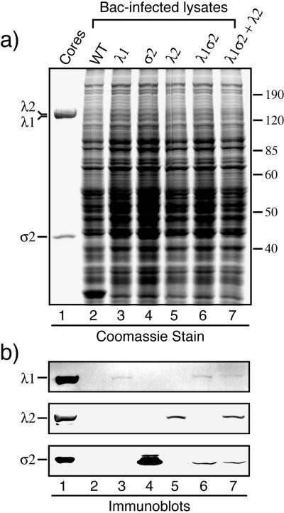
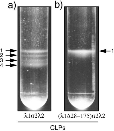
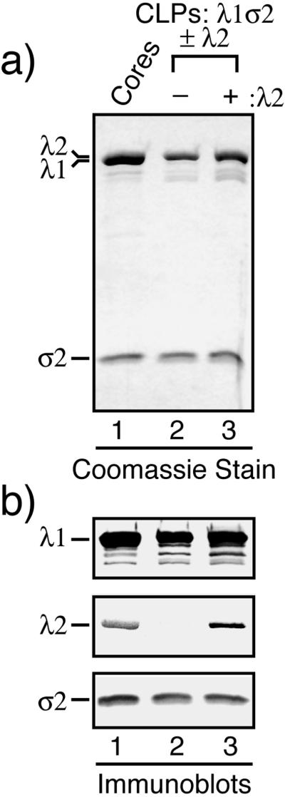
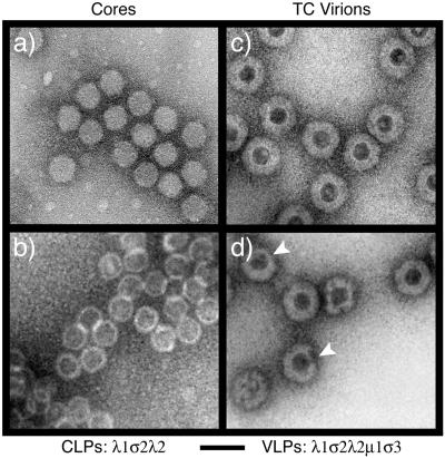
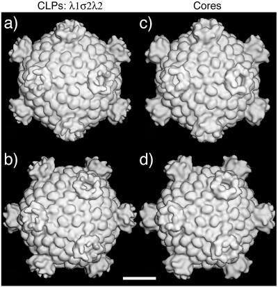
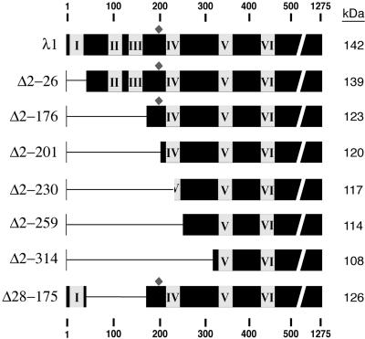
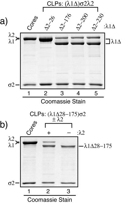
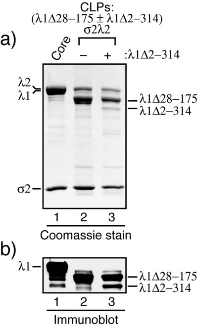
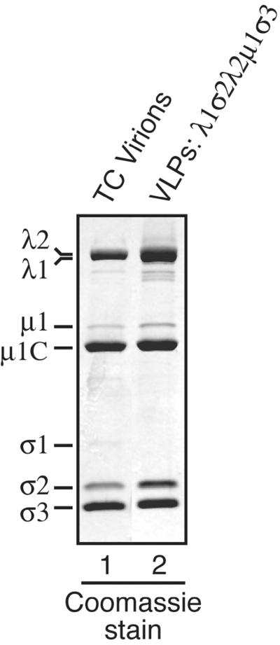
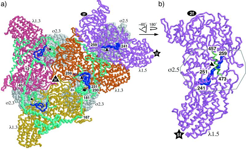
The reovirus core particle is a molecular machine that mediates synthesis, capping, and export of the viral plus strand RNA transcripts. Its assembly and structure-function relationships remain to be well understood. Following the lead of previous studies with other Reoviridae family members, most notably orbiviruses and rotaviruses, we used recombinant baculoviruses to coexpress reovirus core proteins lambda1, lambda2, and sigma2 in insect cells. The resulting core-like particles (CLPs) were purified and characterized. They were found to be similar to cores with regard to their sizes, morphologies, and protein compositions. Like cores, they could also be coated in vitro with the two major outer-capsid proteins, micro 1 and sigma3, to produce virion-like particles. Coexpression of core shell protein lambda1 and core nodule protein sigma2 was sufficient to yield CLPs that could withstand purification, whereas expression of lambda1 alone was not, indicating a required role for sigma2 as a previous study also suggested. In addition, CLPs that lacked lambda2 (formed from lambda1 and sigma2 only) could not be coated with micro 1 and sigma3, indicating a required role for lambda2 in the assembly of these outer-capsid proteins into particles. To extend the use of this system for understanding the core and its assembly, we addressed the hypothesis that the hydrophilic amino-terminal region of lambda1, which adopts an extended arm-like conformation around each threefold axis in the reovirus core crystal structure, plays an important role in assembling the core shell. Using a series of lambda1 deletion mutants, we showed that the amino-terminal 230 residues of lambda1, including its zinc finger, are dispensable for CLP assembly. Residues in the 231-to-259 region of lambda1, however, were required. The core crystal structure suggests that residues in the 231-to-259 region are necessary because they affect the interaction of lambda1 with the threefold and/or fivefold copies of sigma2. An effective system for studies of reovirus core structure, assembly, and functions is hereby established.


Figures

FIG. 1.
FIG. 1.
Expression of the three reovirus core proteins in insect cells using recombinant baculoviruses. S. frugiperda 21 cells were infected with wild-type (WT) baculovirus (lane 2), Bac-λ1L (lane 3), Bac-σ2L (lane 4), Bac-λ2D (lane 5), Bac-λ1Lσ2L (lane 6), or Bac-λ1Lσ2L and Bac-λ2D (lane 7). The cells were harvested at 72 h p.i. and lysed. (a) The soluble cytoplasmic fractions were analyzed by SDS-PAGE and Coomassie staining. Reovirus T1L core particles (lane 1) were included on the gel to mark the positions of the core proteins. All three λ proteins (142 to 144 kDa each) comigrate in one band in this type of gel. (b) The same samples, including core particles, were analyzed in the same respective lanes of another gel by SDS-PAGE and immunoblotting using a polyclonal anti-λ1 serum, anti-λ2 monoclonal antibody 7F4, or a polyclonal anti-core serum to detect σ2. Only those portions of the blot containing the respective proteins are shown.
FIG. 2.
FIG. 2.
Appearance of putative CLP bands in CsCl gradients. S. frugiperda 21 cells were infected with Bac-λ1Lσ2L and Bac-λ2D (a) or Bac-λ1L(Δ28-175)σ2L and Bac-λ2D (b). The cells were harvested at 72 h p.i. and processed. The CLPs putatively contained within the cell lysates were allowed to form discrete bands, visible by eye, on a 1.25- to 1.35-g/cm3 CsCl equilibrium gradient and then photographed. Arrows, numbers of discrete bands visible in each gradient. The upper parts of the tubes have been cropped from these images but contained no visible bands.
FIG. 3.
FIG. 3.
Protein compositions of purified CLPs. CLPs were purified from the lysate of S. frugiperda 21 cells infected with either Bac-λ1Lσ2L (lane 2) or Bac-λ1Lσ2L and Bac-λ2D (lane 3). (a) The CLPs were analyzed by SDS-PAGE and Coomassie staining to estimate their protein contents. Reovirus T1L core particles (lane 1) were included on the gel to mark the positions of the core proteins as well as to provide a standard for the relative intensities of the λ and σ2 bands. (b) The same samples, including core particles, were analyzed in the same respective lanes of another gel by SDS-PAGE and immunoblotting as described for Fig. 1b.
FIG. 4.
FIG. 4.
Transmission electron micrographs of negatively stained reovirus particles. Purified T1L cores (a), λ1σ2λ2 CLPs (b), T1L TC virions (c), and λ1σ2λ2μ1σ3 VLPs (d) were absorbed onto carbon-coated copper grids, stained with 1% uranyl acetate, and observed by EM. Arrowheads (d), partially coated VLPs. Bar, 100 nm.
FIG. 5.
FIG. 5.
3D reconstructions of reovirus particles obtained by image processing of transmission cryoelectron micrographs. Purified λ1σ2λ2 CLPs (a and b) and T1L cores (c and d) were examined. Each reconstruction is shown as a surface-shaded view down either a twofold axis (a and c) or a threefold axis (b and d) at a resolution of 32 Å. Bar, 20 nm.
FIG. 6.
FIG. 6.
Schematic diagram of λ1 deletion mutants. Deletions in the T1L L3 gene were constructed in order to express versions of the full-length T1L λ1 protein (1,275 amino acids, 142 kDa) with deletions. The missing region in each deletion mutant is indicated by the horizontal narrow line. As a result of these deletions, the different versions of λ1 differ in expected molecular mass as indicated at the right. Numbers at the top and bottom are residue numbers. Boxes I to VI, positions of the six putative helicase-like motifs (7, 48); diamonds, positions of the zinc finger motifs (6, 27, 50).
FIG. 7.
FIG. 7.
Protein compositions of purified CLPs formed with the λ1 deletion mutants. CLPs were formed following coexpression of the reovirus core proteins in S. frugiperda 21 cells by infections with Bac-λ1LΔ2-26σ2L, Bac-λ1LΔ2-176σ2L, Bac-λ1LΔ2-200σ2L, Bac-λ1LΔ2-230σ2L, Bac-λ1LΔ28-175σ2L, and/or Bac-λ2D as indicated. The CLPs were then purified from the infected-cell lysates. (a) Each of the indicated λ1 deletion mutants was coexpressed along with σ2 and λ2. The resulting CLPs were analyzed by SDS-PAGE and Coomassie staining. (b) λ1Δ28-175, as a representative of CLP-positive λ1 deletion mutants, was coexpressed with σ2 in either the presence (+) or the absence (−) of λ2. The resulting CLPs were analyzed by SDS-PAGE and Coomassie staining.
FIG. 8.
FIG. 8.
Complementation of a CLP-negative λ1 mutant with a CLP-positive one. One set of S. frugiperda 21 cells was infected to coexpress λ1Δ28-175 (as a representative of the CLP-positive λ1 deletion mutants), σ2, and λ2, while another set of S. frugiperda 21 cells was infected to coexpress both λ1Δ28-175 and λ1Δ2-314λ1 (a CLP-negative mutant) in addition to σ2 and λ2. The two sets of cells were processed in parallel to obtain purified CLPs. The CLPs were then analyzed by SDS-PAGE followed by Coomassie staining (a) or immunoblotting using the polyclonal anti-λ1 serum (b). The bands corresponding to the λ1 deletion mutants are marked at the right.
FIG. 9.
FIG. 9.
Coating CLPs with complexes of the major outer-capsid proteins μ1 and σ3 to generate VLPs. (a) An S. frugiperda 21 cell lysate containing λ1σ2λ2 CLPs was mixed with a lysate of T. ni High Five cells containing complexes of μ1 and σ3 (11, 36). The mixed lysate was incubated and processed to obtain purified particles. These putative VLPs were then analyzed by SDS-PAGE and Coomassie staining (lane 2). Reovirus T1L TC virions (lane 1) were included on the gel to mark the positions of the μ1/μ1C and σ3 proteins as well as to provide a standard for the relative intensities of these bands. The σ1 protein is missing from the VLPs because it was not added in this experiment.
FIG. 10.
FIG. 10.
Views of λ1 and σ2 from the reovirus core crystal structure. Approximate locations of the threefold (▴), twofold (igwidth>), and fivefold (★) axes are marked. (a) Inside-to-outside view of a portion of the T=1 λ1 shell with accompanying σ2 subunits, centered on a threefold axis of the core (50) (see Fig. 5 for whole-particle surface views that aid in orientation). Three λ1.3 subunits are shown, mostly in pink, orange, or yellow (backbone format). One λ1.5 subunit is also shown, mostly in violet (backbone format). The σ2.3 and σ2.5 subunits that associate with the tops of these λ1 subunits are shown in light gray (space-filling format). Amino acids 1 to 259 of each λ1 subunit (maximum extent of N-terminal sequences that were removed from the deletion mutants in this study) are shown in cyan or blue for emphasis. Amino acids 1 to 240 were not visualized in the core crystal structure of the λ1.5 subunits (50) and are thus not shown in this diagram. In the one λ1.5 subunit pictured, amino acids 241 to 259 are shown in blue as well as in thicker backbone format for greater emphasis. The 251-to-259 region of this subunit adopts an extended conformation and is seen diving into the main body of λ1 (arrowhead) beneath the σ2.5 subunit. Amino acids 1 to 259 of one λ1.3 subunit (the one for which amino acids 260 to 1275 are shown in orange) are shown in thicker backbone format for greater emphasis of that subunit. Amino acids 13 to 39 and 168 to 180 were not visualized in the core crystal structure of the λ1.3 subunits (50), but their proposed locations (50) are shown for the emphasized λ1.3 subunit as straight lines between the flanking visualized regions. In all three λ1.3 subunits pictured, amino acids 1 to 230 are shown in cyan and amino acids 231 to 259 are shown in blue. The 251-to-259 regions of these subunits also adopt extended conformations and can be seen diving into the main body of λ1 (arrowhead for the emphasized subunit) beneath the σ2.3 subunit. The zinc ion in the zinc finger of each λ1.3 subunit is shown as a black ball, which is larger and labeled for the emphasized subunit. (b) The violet λ1.5 subunit from panel a was enlarged and rotated as indicated such that the view of this subunit is now that from outside the core. Amino acids 241 to 259 are shown in blue and in thicker backbone format as in panel a. The arrowhead points to approximately the same position in the λ1.5 subunit as in panel a. The α-helix composed of amino acids 457 to 473, which overlies the 251-to-259 region, is shown in light and dark green. Residues 463, 464, 467, 470, and 471, which make extensive contacts with the base of the overlying σ2.5 subunit, are shown in dark green. Only the outline of the σ2.5 subunit is shown to allow clear views of the underlying λ1.5 subunit.

Similar articles

Cited by

References

    1. Acs, G., H. Klett, M. Schonberg, J. Christman, D. H. Levin, and S. C. Silverstein. 1971. Mechanism of reovirus double-stranded ribonucleic acid synthesis in vivo and in vitro. J. Virol. 8:684-689. - PMC - PubMed
    1. Baker, T. S., and R. H. Cheng. 1996. A model-based approach for determining orientations of biological macromolecules imaged by cryo-electron microscopy. J. Struct. Biol. 116:120-130. - PubMed
    1. Baker, T. S., N. H. Olson, and S. D. Fuller. 1999. Adding the third dimension to virus life cycles: three-dimensional reconstruction of icosahedral viruses from cryo-electron micrographs. Microbiol. Mol. Biol. Rev. 63:862-922. - PMC - PubMed
    1. Banerjee, A. K., and M. A. Grece. 1971. An identical 3′-terminal sequence in the ten reovirus genome RNA segments. Biochem. Biophys. Res. Commun. 45:1518-1525. - PubMed
    1. Banerjee, A. K., R. Ward, and A. J. Shatkin. 1971. Cytosine at the 3′-termini of reovirus genome and in vitro mRNA. Nat. New Biol. 232:114-115. - PubMed

Publication types

MeSH terms

LinkOut - more resources