. 2002 Dec;76(23):12250-8.
doi: 10.1128/jvi.76.23.12250-12258.2002.

An antibody to the putative aphid recognition site on cucumber mosaic virus recognizes pentons but not hexons

Affiliations

An antibody to the putative aphid recognition site on cucumber mosaic virus recognizes pentons but not hexons

Valorie D Bowman et al. J Virol. 2002 Dec.

Abstract

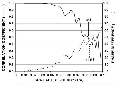
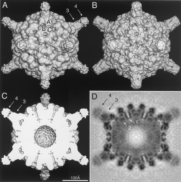
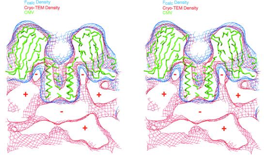
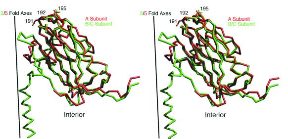
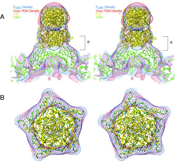
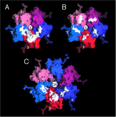
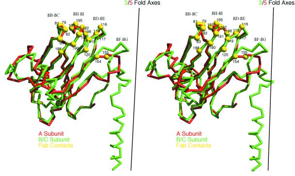
Cucumber mosaic virus (CMV), the type member of the genus Cucumovirus (family Bromoviridae), is transmitted by aphids in a nonpersistent manner. Mutagenesis experiments identified the betaH-betaI loop of the capsid subunit as a potential key motif responsible for interactions with the insect vector. To further examine the functional characteristics of this motif, we generated monoclonal antibodies that bound to native virions but not to betaH-betaI mutants. Fab fragments from these antibodies were complexed with wild-type CMV and the virus-Fab structure was determined to 12-A resolution by using electron cryomicroscopy and image reconstruction techniques. The electron density attributed to the bound antibody has a turret-like appearance and protrudes from each of the 12 fivefold axes of the icosahedral virus. Thus, the antibody binds only to the pentameric clusters (pentons) of A subunits of the T=3 quasisymmetric virus and does not appear to bind to any of the B and C subunits that occur as hexameric clusters (hexons) at the threefold (quasi-sixfold) axes. Modeling and electron density comparisons were used to analyze the paratope-epitope interface and demonstrated that the antibody binds to three betaH-betaI loops in three adjacent A subunits in each penton. This antibody can discriminate between A and B/C subunits even though the betaH-betaI loop adopts the same structure in all 180 capsid subunits and is therefore recognizing differences in subunit arrangements. Antibodies with such character have potential use as probes of viral assembly. Our results may provide an additional rationale for designing synthetic vaccines by using symmetrical viral particles.


Figures

FIG. 1.
FIG. 1.
Resolution assessment of CMV-Fab 3D reconstruction. The final set of boxed CMV-Fab images was subdivided in half, and two independent reconstructions were computed. Correlation coefficient (solid line) and phase difference (dashed line) comparisons were computed from the structure factors derived from the two reconstructions. Based on conservative estimates (CC, >0.5; phase differences, <45°), these plots demonstrate that the data are reliable to at least 12-Å resolution, and significant Fourier terms are present to ca. 10.5 Å.
FIG. 2.
FIG. 2.
Shaded-surface (A to C) and equatorial section (D) representations of the CMV-Fab complex, all viewed along a twofold axis of symmetry. The arrows highlight aspects of the cryoTEM 3D reconstruction that are depicted in greater detail in subsequent figures. (A) 3D reconstruction of the CMV-Fab complex with the approximate locations of the three quasiequivalent (chemically identical) A, B, and C subunits labeled. The large, turret-like projections at the icosahedral vertices represent the bound Fab molecules, and the variable and constant domains of one Fab are identified by arrows 3 and 4, respectively. (B) Pseudo-atomic model of CMV-Fab complex computed to 10-Å resolution; β factors were applied to match the 12-Å cryo-TEM data, using the X-ray crystal structures of CMV (32) and Fab17 (19). (C) Same as panel A with the front half of the structure removed to visualize internal features of the reconstruction. (D) Density projection of the central section of the CMV-Fab reconstruction, with the blackest regions representing the highest density regions in the map. Two key internal features are the radially directed density representing the six-helical bundle at the quasi-sixfold axes (arrow 1) and density that lies adjacent to and projects away from the helical bundle (arrow 2). The lower density for the Fabs (especially the constant domains [arrow 4]) is consistent with the hypothesis that only a single Fab binds to each of the 12 pentons at the vertices of the icosahedral virus.
FIG. 3.
FIG. 3.
Stereo cross-section view of CMV density maps and X-ray model in region near a quasi-sixfold axis. The 3D reconstruction and X-ray map (rendered at a 15-Å resolution) are depicted in red and blue contours, respectively. The C-α backbone of the CMV model is represented by thin green tubes. Selected regions of positive (high) and negative (low or zero) cryoTEM electron density are denoted by “+” and “−” signs for clarity. Two large regions of positive density stream away from the sides of the helical bundle (see also arrow 2, Fig. 2). Negative density, centered directly beneath the helical bundle, occurs in a region that contains positive density in CCMV and which is attributed to organized, genomic RNA (37). Figures 3 to 5 and 7 were made by using the program MolView (http://www.danforthcenter.org/smith/molview.htm [31]).
FIG. 4.
FIG. 4.
Stereo view comparison of the tertiary structures of the A (red) and B/C (green) subunits in CMV. The subunits are aligned with the icosahedral three- and fivefold axes superimposed. The RNA core of the virus (not shown) starts at the bottom of the panel. The side chains for residues 191, 192, and 195 are depicted to illustrate the conservation of main and side chain conformations, especially at the viral surface of the quasiequivalent subunits.
FIG. 5.
FIG. 5.
Stereo view showing fit of the CMV (green) and Fab (yellow) X-ray models into the cryoTEM electron density map (red contours) and the electron density calculated from the pseudo-atomic model (blue contours). A single Fab was fit into the reconstructed density envelope, and then icosahedral fivefold symmetry was applied. Since the density envelope could only accommodate one Fab at each penton, an occupancy of 0.2 was applied to each of the fivefold-related Fab molecules. Therefore, although all five Fabs are shown in this figure to demonstrate how the cryoTEM electron density is filled by the Fab structures, in reality only one Fab molecule is bound at a time. (A) Close-up view of the CMV-Fab model and density maps, with the viral RNA core at the bottom of the figure and a fivefold axis aligned in a vertical direction. (B) Same as panel A but viewed down a fivefold axis and including just a portion of the “turret” corresponding to the region demarked by brackets labeled “B” in panel A. Agreement between the calculated and observed maps is excellent. Also, the averaged variable domains of the Fabs form a pentameric structure docked to the surface of the A subunit penton.
FIG. 6.
FIG. 6.
Molecular surface representations of CMV penton (A and B) and hexon (C) with Fab contact area and location of mutations depicted. (A) Contact area (white) of a single Fab on a penton with individual subunits distinguished by different colors and the four subunits noted in Table 1 are labeled 1 to 4. The view is along the fivefold axis toward the virus center. A single Fab contacts significant regions of three different A subunits. (B) Same as panel A but with white coloring to denote the locations and surface formed by residues, 191, 192, and 195. Based on panel A, the Fab is seen to contact some or all of these residues in penton subunits 2, 3, and 4. (C) Mapping of contact residues seen in panel A onto the hexon B and C subunits demonstrates that the hypothetical hexon contact is discontinuous compared to the contact observed in the penton. This figure was made by using the program GRASP (24).
FIG. 7.
FIG. 7.
Stereo view of aligned A (red) and B/C (green) subunit Cα backbones with contact residues (yellow) within a penton mapped onto a single A subunit. The three major contact loops (βB-βC, βH-βI, and βD-βE) have nearly identical structures in the quasiequivalent subunits (see also Fig. 4). Some contact with the βF-βG loop, which bends away from the fivefold axis in the A subunit, may occur, but this loop only contributes ∼3% of the total virus-Fab contact area and therefore may not represent an actual contact in the real virus-antibody complex.

Similar articles

Cited by

References

    1. Bailey, S. 1994. The CCP4 suite: programs for protein crystallography. Acta Crystallogr. D 50:760-763. - PubMed
    1. Baker, T. S., and R. H. Cheng. 1996. A model-based approach for determining orientations of biological macromolecules imaged by cryoelectron microscopy. J. Struct. Biol. 116:120-130. - PubMed
    1. Baker, T. S., N. H. Olson, and S. D. Fuller. 1999. Adding the third dimension to virus life cycles: three-dimensional reconstruction of icosahedral viruses from cryo-electron micrographs. Microbiol. Mol. Biol. Rev. 63:862-922. - PMC - PubMed
    1. Booy, F. P., R. B. S. Roden, H. L. Greenstone, J. T. Schiller, and B. L. Trus. 1998. Two antibodies that neutralize papillomavirus by different mechanisms show distinct binding patterns at 13 Å resolution. J. Mol. Biol. 281:95-106. - PubMed
    1. Burke, K. L., G. Dunn, M. Ferguson, P. Minor, and J. W. Almond. 1988. Antigen chimeras of poliovirus as potential vaccines. Nature 332:81-82. - PubMed

Publication types

MeSH terms

LinkOut - more resources