. 2002 Dec;76(23):12325-34.
doi: 10.1128/jvi.76.23.12325-12334.2002.

Generation of hepatitis C virus-like particles by use of a recombinant vesicular stomatitis virus vector

Affiliations

Generation of hepatitis C virus-like particles by use of a recombinant vesicular stomatitis virus vector

Heather J Ezelle et al. J Virol. 2002 Dec.

Abstract

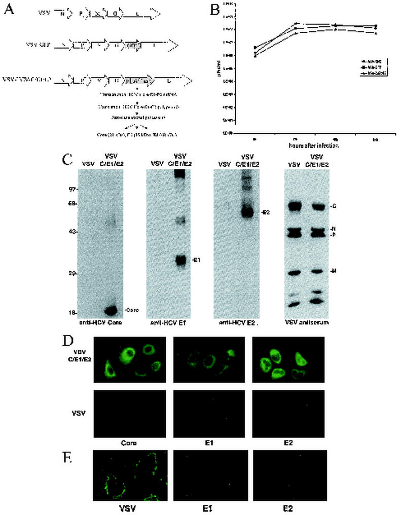
Hepatitis C virus (HCV), a major etiologic agent of hepatocellular carcinoma, presently infects approximately 400 million people worldwide, making the development of protective measures against HCV infection a key objective. Here we have generated a recombinant vesicular stomatitis virus (VSV), which expresses the HCV structural proteins, by inserting the contiguous Core, E1, and E2 coding region of HCV into the VSV genome. Recombinant VSV expressing HCV Core, E1, and E2 (VSV-HCV-C/E1/E2) grew to high titers in vitro and efficiently expressed the incorporated HCV gene product, which became fully processed into the individual HCV structural proteins. Biochemical and biophysical analysis indicated that the HCV Core, E1, and E2 proteins assembled to form HCV-like particles (HCV-LPs) possessing properties similar to the ultrastructural properties of HCV virions. Mice immunized with VSV-HCV-C/E1/E2 generated cell-mediated immune responses to all of the HCV structural proteins, and humoral responses, particularly to E2, were also readily evident. Our data collectively indicate that engineered VSVs expressing HCV Core, E1, and E2 and/or HCV-LPs represent useful tools in vaccine and immunotherapeutic strategies designed to address HCV infection.


Figures

FIG. 1.
FIG. 1.
(A) Construction of rVSV expressing HCV Core, E1, and E2. The Core, E1, and E2 regions (aa 1 to 746) of the HCV polypeptide (NIHJ1 provided by T. Miyamura) were cloned into the XhoI and NheI sites of the rVSV replicon vector pVSV-XN2 (provided by J. Rose) by PCR using the forward primer 5′-CTCGTAGCTCGAGCATCATGAGCACAAATC-3′ and the reverse primer 5′-ACCAAGTTCTCTAGA CTAAGCCTCGGCCTGGGCTAT-39. Recovery of rVSV and the construction of VSV-GFP have been previously described (23). (B) Growth-Growth analysis of recombinant viruses. VSV-HCV-C/E1/E2 demonstrates a growth rate similar to that of rVSV-GFP. BHK cells were infected at an MOI of 1 for 30 min. One hundred microliters of cell medium was collected at 6, 12, 18, and 24 h postinfection and virus titers were determined by plaque assay, as described previously (4). (C) Expression of HCV Core, E1, and E2. BHK cells were infected with VSV-HCV-C/E1/E2 or control virus VSV-XN2 at an MOI of 1. After 18 h, cells were lysed and HCV protein expression was determined by immunoblot analysis as previously described (19). HCV proteins were detected by anti-Core MAb (Biogenesis, Poole, United Kingdom), anti-E1 (a gift from S. J. Polyak), and anti-E2 (WU105; a gift from C. M. Rice). VSV proteins were detected by polyclonal mouse antiserum generated in BALB/c mice infected with VSV. (D) Immunofluorescence analysis of HCV structural proteins. Expression and intracellular localization of HCV structural proteins were confirmed by immunofluorescence using MAbs specific for Core (Biogenesis), E1 (A4; a gift from H. Greenberg), or E2 (544; a gift from M. Kohara). Briefly, Huh-7 cells (provided by S. Lemon) were infected with VSV-XN2 or VSV-HCV-C/E1/E2 at an MOI of 10 for 5 h and were then fixed in 1% paraformaldehyde. The cells were incubated in 1:50 dilutions of primary antibody in 0.1% Brij 97_PBS for 2 h at 4°C and were then incubated with fluorescein isothiocyanate-conjugated goat anti-mouse (1:100; Gibco-BRL, Grand Island, N.Y.) in 0.1% Brij 97_PBS for 1 h at 4°C. (E) Cell surface immunofluorescence staining of VSV-C/E1/E2-infected cells. Immunofluorescence analysis of infected Huh-7 cells was performed as for panel D except that the cells were not permeabilized with Brij 97. VSV-HCV-C/E1/E2-infected cells were stained for cell surface expression of E1 or E2 using MAbs or for expression of VSV using polyclonal mouse serum.
FIG. 2.
FIG. 2.
(A and B) HCV E2 is not associated with VSV-HCV-C/E1/E2. Cell medium from VSV-HCV-C/E1/E2- or control virus-infected cells (concentrated by ultracentrifugation) was immunoprecipitated (IP) with a sheep antibody (Ab) to VSV G (Biogenesis) or a goat antibody to HCV E2 (Immunodiagnostics Inc.). After SDS-PAGE, protein complexes were immunoblotted against mouse antiserum raised to VSV (A). Membranes were reprobed using mouse antiserum to HCV E2 (M. Kohara), which emphasized the absence of E2 in VSV complexes (B). cntl represents protein G agarose, which was used as a negative control. (C) Gradient-purified HCV Core, E1, and E2 form complexes. Sucrose gradient fractions containing HCV-LPs were identified by immunoblot and were immunoprecipitated using E2-specific antibody (Immunodiagnostics Inc.). Complexes were washed, analyzed by SDS-PAGE, and immunoblotted using antibody to HCV Core (Biogenesis), E1 (H. Greenberg), and E2 (M. Kohara). (D) Coimmunoprecipitation analysis of VSV-HCV-C/E1/E2-infected cells. [35S]methionine/cysteine-labeled lysates (600 μCi/12 h) from mock, VSV-XN2, or VSV-HCV-C/E1/E2 were immunoprecipitated with mouse antiserum raised to VSV or anti-E2 MAb (544; M. Kohara) or normal mouse immunoglobulin G. Complexes were washed, analyzed by SDS-PAGE, and visualized by autoradiography. (E) Analysis of HCV-LPs by sucrose gradients. BHK cells were infected at an MOI of 0.1 for 18 h and were then lysed in 50 mM Tris-HCl, pH 7.5, 50 mM NaCl, 0.1% NP-40, 1 mM phenylmethylsulfonyl fluoride, 10 μg of aprotinin/ml, 10 μg of leupeptin/ml, and 0.5 mM EDTA. The lysates were clarified by centrifugation through 30% sucrose for 6 h at 150,000 × g. The resulting pellets were layered onto a continuous 30 to 70% sucrose gradient. One-milliliter fractions were collected, centrifuged, and analyzed by SDS-PAGE and immunoblotting using antibody to VSV (top gel) or HCV Core (Biogenesis), E1 (S. Polyak), or E2 (C. Rice) (bottom three gels). HCV structural proteins colocalize in fractions 14 to 20 and are separable from VSV proteins.
FIG. 3.
FIG. 3.
(A to D) Electron microscopy images of BHK cells infected with VSV-HCV-C/E1/E2. BHK cells were mock infected or infected at an MOI of 0.1 with VSV-XN2 or VSV-HCV-C/E1/E2 for 18 h. Cells were fixed in 2% paraformaldehyde-2.5% glutaraldehyde and were incubated in 1% osmium tetroxide for 1 h. Fixed cells were then dehydrated and embedded in Spurr's resin. Thin sections were stained in aqueous 4% uranyl acetate for 20 min followed by lead citrate. (A) Mock- infected BHK cells. Magnification is ×19,444. (B) VSV-XN2-infected BHK cells producing the characteristically bullet-shaped VSV virion. Magnification is ×55,163. Bar is 100 nm. (C) VSV-HCV-C/E1/E2-infected BHK cells at ×19,772 magnification. Black arrows indicate HCV-LPs in vacuoles formed from rough ER. (D) Higher magnification of the vacuole containing HCV-LPs in panel C. Asterisk in panel C indicates the magnified vacuole. Bar is 100 nm (magnification is ×44,190). (E to H) Electron microscopy images of HCV-LPs purified by equilibrium sedimentation sucrose gradients. Gradient-purified particles were adsorbed to carbon-coated copper grids and were then negative stained with 2% uranyl acetate for 2 or 3 min (E) (magnification is ×77,333). (F to H) For immunogold labeling, HCV-LPs were incubated with anti-E1 MAb, magnification, ×259,200 (F) (A4; H. Greenberg); anti-E2 MAb, magnification, ×171,428 (G) (544; M. Kohara); or control mouse immunoglobulin G, magnification, ×214,050 (H), and were stained with 2% uranyl acetate. Bar in inset of panel G is 100 nm.
FIG. 4.
FIG. 4.
Humoral and cellular immune responses generated to HCV structural proteins using VSV as a vector. (A) Humoral activity to HCV E2. ELISA plates (Nunc) adsorbed with E2 protein were used to detect E2 antibody production following i.v. immunization with VSV-HCV-C/E1/E2 but not with VSV-GFP- or PBS-treated control mice (six mice each group) at a dilution of 1/100. Each bar represents a pool of three mice. (B) Humoral activity to HCV E2 generated by i.p. immunization. Anti-E2 ELISAs were conducted on serum collected from mice vaccinated i.p. with VSV-HCV-C/E1/E2, VSV-GFP, or PBS. Each bar represents a single mouse at a dilution of 1/100. OD, optical density. (C) Lymphoproliferative response to HCV. Single-cell preparations from spleens of i.v. immunized mice were suspended in RPMI 1640 supplemented with 10% fetal bovine serum (Sigma, St. Louis, Mo.), 1 mM sodium pyruvate, 100 U of penicillin per ml, and 100 μg of streptomycin (Gibco-BRL) per ml. The cells were stimulated with recombinant HCV core protein (1, 0.1, and 0.01 μg/ml) (Advanced Immunochemical, Long Beach, Calif.). As a negative control, spleen cells were stimulated with medium alone. The wells were pulsed with 1 μCi of [3H]thymidine per well for the last 18 h of a 5-day period. The results are expressed as mean values of triplicate determinations. (D) CTL activation by HCV Core, E1, and E2. IFN-γ production was determined by ELISPOT analysis as previously described (1). Cells harvested from the spleens of i.v. immunized animals (VSV-HCV-C/E1/E2, VSV-GFP, or PBS) were placed in wells in triplicate using four different cell concentrations in twofold dilutions (from 106). These results are representative of three independent experiments, each utilizing a minimum of two mice per group. Each peptide location is noted below the respective HCV protein. (E) IFN-γ ELISPOT analysis of i.p. vaccinated mice. Mice vaccinated i.p. were analyzed for IFN-γ production 7 days following the injection. The results indicate that only VSV-HCV-C/E1/E2-vaccinated mice activate T cells when pulsed with HCV peptides.

Comment in

Similar articles

Cited by

References

    1. Adkins, B., Y. Bu, and P. Guevara. 2001. The generation of Th memory in neonates versus adults: prolonged primary Th2 effector function and impaired development of Th1 memory effector function in murine neonates. J. Immunol. 166:918-925. - PubMed
    1. Aizaki, H., Y. Aoki, T. Harada, K. Ishii, T. Suzuki, S. Nagamori, G. Toda, Y. Matsuura, and T. Miyamura. 1998. Full-length complementary DNA of hepatitis C virus genome from an infectious blood sample. Hepatology 27:621-627. - PubMed
    1. Arichi, T., T. Saito, M. E. Major, I. M. Belyakov, M. Shirai, V. H. Engelhard, S. M. Feinstone, and J. A. Berzofsky. 2000. Prophylactic DNA vaccine for hepatitis C virus (HCV) infection: HCV-specific cytotoxic T lymphocyte induction and protection from HCV-recombinant vaccinia infection in an HLA-A2.1 transgenic mouse model. Proc. Natl. Acad. Sci. USA 97:297-302. - PMC - PubMed
    1. Balachandran, S., P. C. Roberts, T. Kipperman, K. N. Bhalla, R. W. Compans, D. R. Archer, and G. N. Barber. 2000. Alpha/beta interferons potentiate virus-induced apoptosis through activation of the FADD/caspase-8 death signaling pathway. J. Virol. 74:1513-1523. - PMC - PubMed
    1. Baumert, T. F., S. Ito, D. T. Wong, and T. J. Liang. 1998. Hepatitis C virus structural proteins assemble into viruslike particles in insect cells. J. Virol. 72:3827-3836. - PMC - PubMed

MeSH terms

Substances

LinkOut - more resources