. 2002 Nov 12;99(23):15042-7.
doi: 10.1073/pnas.232588799. Epub 2002 Nov 1.

Down-regulation of parkin protein in transient focal cerebral ischemia: A link between stroke and degenerative disease?

Affiliations

Down-regulation of parkin protein in transient focal cerebral ischemia: A link between stroke and degenerative disease?

Thorsten Mengesdorf et al. Proc Natl Acad Sci U S A. .

Abstract

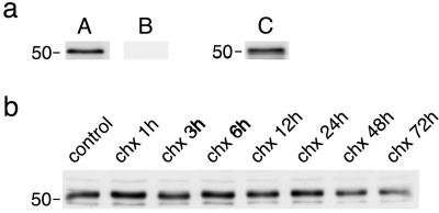
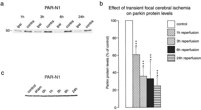
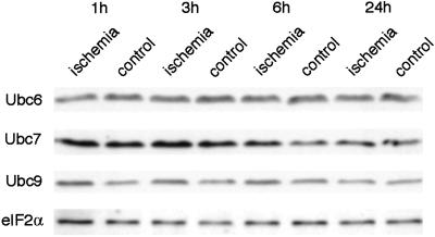
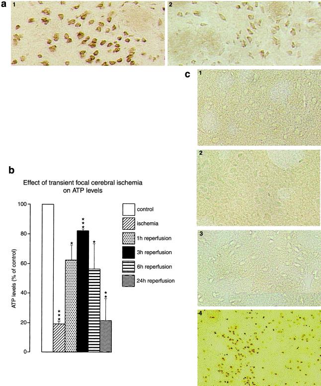
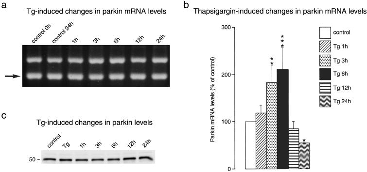
Ubiquitylated protein aggregates are characteristic features of neurodegenerative disorders that are also found in acute pathological states of the brain such as stroke. Many of the proteins connected to neurodegenerative diseases play a role in the ubiquitin-proteasomal pathway. Mutation of one of these proteins, the E3 ubiquitin ligase parkin, is the cause of autosomal recessive juvenile Parkinson's disease. Here we show that transient focal cerebral ischemia of 1-h duration induces marked depletion of parkin protein levels, to 60%, 36%, 33%, and 25% of controls after 1, 3, 6, and 24 h of reperfusion, but that ischemia does not cause lower protein levels of E2 ubiquitin-conjugating enzymes Ubc6, Ubc7, or Ubc9. After 3 h of reperfusion, when parkin protein levels were already reduced to <40% of control, ATP levels were almost completely recovered from ischemia and we did not observe DNA fragmentation, suggesting that parkin depletion preceded development of neuronal cell death. Up-regulation of the expression of parkin has been shown to protect cells from injury induced by endoplasmic reticulum (ER) dysfunction, and this form of cellular stress is also triggered by transient cerebral ischemia. However, in contrast to observations in neuroblastoma cells, we saw no up-regulation of parkin expression in primary neuronal cell cultures after induction of ER dysfunction. Our data thus suggest that ischemia-induced depletion of parkin protein may contribute to the pathological process resulting in cell injury by increasing the sensitivity of neurons to ER dysfunction and the aggregation of ubiquitylated proteins during the reperfusion period.


Figures

Fig 1.
Fig 1.
Immunoblotting of parkin protein. Protein extracts from control brains were used to evaluate parkin immunoblotting with the PAR-N1 and T160 antibody. (a) A protein band of ≈52 kDa was present in immunoblots produced with the PAR-N1 antibody (A). Appearance of the band was blocked when the PAR-N1 antibody was preincubated with the PAR-N1 peptide (B), and the band also appeared in immunoblots prepared with the T160 antibody (C). (b) Changes in 52-kDa parkin band after blocking protein synthesis by exposing primary neuronal cell cultures to cycloheximide (chx; 20 μg/ml) for the indicated periods of time.
Fig 2.
Fig 2.
Ischemia-induced changes in parkin protein levels. Mice were subjected to 1 h of left MCA occlusion, followed by varying periods of reperfusion. In samples derived from the ischemic left MCA territory, the parkin band started to disappear after 1 h of reperfusion (a). Quantification of ODs of the parkin band revealed a marked decrease of parkin protein levels (b; ***, P > 0.001, left versus right hemisphere). In the contralateral nonischemic hemisphere, parkin protein levels did not change during ischemia or after reperfusion (c). Western blots were run with samples from the MCA territory of plain control animals, sham-operated animals, and the nonischemic MCA territory of the right hemisphere of animals subjected to left MCA occlusion without recovery (0 h) or with 1–24 h of reperfusion.
Fig 3.
Fig 3.
Ischemia-induced changes in Ubc6, Ubc7, and Ubc9 protein levels and levels of eIF2α. Ischemia-induced changes in Ubc6, Ubc7, Ubc9, and eIF2α protein levels were evaluated after 1 h of MCA occlusion followed by the indicated reperfusion times. Transient focal cerebral ischemia did not cause a decrease but caused a transient increase in protein levels of ubiquitin-conjugating enzymes Ubc7 and Ubc9. Protein levels of Ubc6 and eIF2α did not change after transient focal cerebral ischemia.
Fig 4.
Fig 4.
Ischemia-induced changes in neuronal parkin protein levels (a, immunohistochemistry, 3 h of reperfusion), ATP levels in the territory supplied by the left MCA (b), and DNA fragmentation (c). (a) Parkin immunohistochemistry was performed by using the PAR-N antibody: 1, contralateral striatum; 2, ipsilateral striatum (×200). (c) DNA fragmentation was evaluated by using the TUNEL technique: 1, control; 2, 1 h of reperfusion; 3, 3 h of reperfusion; 4, 72 h of reperfusion. Focal cerebral ischemia caused an almost complete depletion of ATP levels. ATP levels recovered transiently on onset of reperfusion, peaking at 3 h of restoration of blood flow. After 72 h of reperfusion, most of the neurons of the ipsilateral MCA territory showed massive brown deposits, indicating DNA double-strand breaks. After 1 or 3 h of reperfusion, however, we did not observe TUNEL-positive cells, indicating the absence of DNA double-strand breaks (×200).
Fig 5.
Fig 5.
Tg-induced changes in parkin expression. Primary neuronal cell cultures were exposed to Tg (1 μM) or the Tg solvent DMSO (0.1%) for 30 min, followed by varying recovery times in Tg-free medium. PCR products were separated on 2% agarose gels (a). The internal standard band is highlighted by an arrow. Gels were photographed and OD of bands was analyzed by image analysis (b). *, P < 0.05; **, P < 0.01; ***, P < 0.001 (ANOVA, followed by Fisher's protected least-significant difference test). (c) Tg-induced changes in parkin protein levels.

References

    1. Kleihues P., Hossmann, K.-A., Pegg, A. E., Kobayashi, K. & Zimmermann, V. (1975) Brain Res. 95, 61-73. - PubMed
    1. Cooper H. K., Zalewska, T., Kawakami, S. & Hossmann, K.-A. (1977) J. Neurochem. 28, 929-934. - PubMed
    1. Burda J., Martin, M. E., García, A., Alcázar, A., Fando, J. L. & Salinas, M. (1994) Biochem. J. 302, 335-338. - PMC - PubMed
    1. Kumar R., Azam, S., Sullivan, J. M., Owen, C., Cavener, D. R. C., Zhang, P., Ron, D., Harding, H. P., Chen, J.-J., Han, A., et al. (2001) J. Neurochem. 77, 1418-1421. - PubMed
    1. Harding H. P., Zhang, Y. & Ron, D. (1999) Nature 397, 271-274. - PubMed

Publication types

MeSH terms