. 2002 Dec;3(12):1135-41.
doi: 10.1038/ni852. Epub 2002 Nov 4.

Langerhans cells renew in the skin throughout life under steady-state conditions

Affiliations

Langerhans cells renew in the skin throughout life under steady-state conditions

Miriam Merad et al. Nat Immunol. 2002 Dec.

Erratum in

  • Nat Immunol 2003 Jan;4(1):92

Abstract

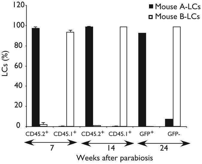
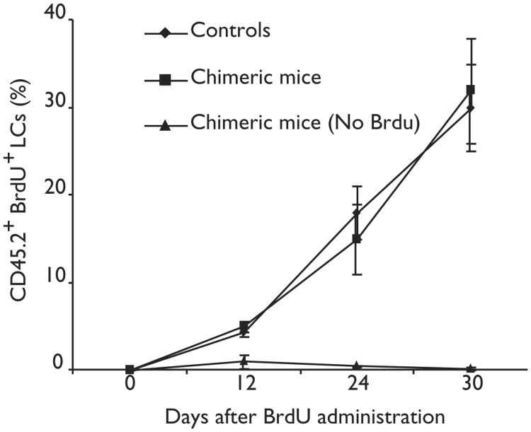
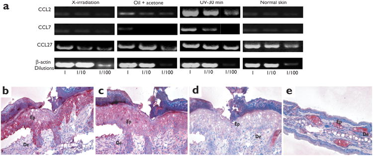
Langerhans cells (LCs) are bone marrow (BM)-derived epidermal dendritic cells (DCs) that represent a critical immunologic barrier to the external environment, but little is known about their life cycle. Here, we show that in lethally irradiated mice that had received BM transplants, LCs of host origin remained for at least 18 months, whereas DCs in other organs were almost completely replaced by donor cells within 2 months. In parabiotic mice with separate organs, but a shared blood circulation, there was no mixing of LCs. However, in skin exposed to ultraviolet light, LCs rapidly disappeared and were replaced by circulating LC precursors within 2 weeks. The recruitment of new LCs was dependent on their expression of the CCR2 chemokine receptor and on the secretion of CCR2-binding chemokines by inflamed skin. These data indicate that under steady-state conditions, LCs are maintained locally, but inflammatory changes in the skin result in their replacement by blood-borne LC progenitors.


Figures

Figure 1
Figure 1. LC chimerism after congenic BM transplantation
Eight-week-old CD45.2-expressing C57BL/6 mice were lethally irradiated and injected intravenously with 5 × 10⁵ BM cells isolated from congenic CD45.1-expressing mice. (a) Contour plots show the expression of CD45.1 and CD45.2 on gated I-Ab+CD11c+ spleen and kidney DCs, CD11b+Gr-1B220NK1.1 peritoneal macrophages and CD11b+Gr-1 blood monocytes isolated 8 weeks after transplantation. The percentages of CD45.1+- and CD45.2+-gated cells are indicated. (b,c) Epidermal cells were isolated before and at different times after congenic BM transplantation; three to six mice were analyzed independently for each time point. (b) One representative experiment showing the expression of CD45.1 and CD45.2 on gated I-Ab+CD11c+ epidermal LCs. (c) Bar graphs show the mean percentages of LCs that were donor (CD45.1)– or host (CD45.2)–derived at different times after transplantation. (d–f) Epidermal sheets were obtained from (d) nonreconstituted (naïve) C57BL/6 mice or (e,f) chimeric animals 18 months after congenic BM transplantation and stained with (d,e) CD45.2 or (f) CD45.1 mAbs. (g) DC chimerism in inguinal (skin-draining), mesenteric and spleen LNs was determined at 6 months after congenic BM transplantation in four different mice. Bar graphs show the percentages of I-Ab+CD11c+ DCs that expressed CD45.2 in different secondary lymphoid organs in each reconstituted animal.
Figure 2
Figure 2. Origin of LCs in parabiotic mice
Parabiotic mice consisted of one CD45.1+ (mouse A) and one CD45.2+ mouse (mouse B) or one green fluorescent protein (GFP)–transgenic mouse (GFP+, mouse A) and one non-GFP–transgenic mouse (GFP,mouse B) sharing the same blood circulation. Bar graphs show the mean percentages of gated LCs that were CD45.1+ and CD45.2+ in recipient parabiotic mice 7 and 14 weeks after parabiosis or GFP+ and GFP in recipient parabiotic mice 6 months after parabiosis. Data represent the means of two separate experiments.
Figure 3
Figure 3. Rate of LC proliferation in chimeric mice
Groups of five CD45.2+ C57BL/6 mice were reconstituted with congenic CD45.1+ BM. Sixteen weeks later, reconstituted and nonreconstituted CD45.2+ mice (controls) received BrdU in their drinking water for 4 weeks.The percentage of CD45.2+ (host) LCs that had incorporated BrdU was determined by flow cytometry at different times after BrdU administration.
Figure 4
Figure 4. LC homeostasis during skin inflammation
CD45.2+ mice were lethally irradiated and reconstituted with 5 × 10⁵ CD45.1+ BM cells and either left untreated or exposed 8 weeks later to UV light for 10 or 30 min or to a mixture of oil and acetone applied to each ear. (a,b) Three ears from three different animals were collected at different times after skin injury and analyzed to determine (a) the total number of LCs per ear and (b) the percentage of epidermal cells that were host LCs. (c) Dot plots show the expression of CD45.1 and CD45.2 on gated I-Ab+CD11c+PI LCs 3 weeks after x-irradiation alone or x-irradiation followed by skin irritation or UV exposure. (d) Bar graphs show the mean percentages of total epidermal cells that were host- or donor-derived LCs analyzed 3 weeks after skin injury in three different animals. (e–g) Images of epidermal sheets obtained from C57BL/6 animals reconstituted with CD45.1+ BM 12 weeks after exposure to UV for 30 min and stained with (e) CD45.1, (f) I-Ab or (g) CD45.2 mAbs. Magnification: ×20.
Figure 5
Figure 5. Role of CCR2 in the recruitment of circulating LC precursors to inflamed skin
(a) cDNA was obtained from various samples and analyzed by PCR for the expression of CCR2. Blood I-Ab−CD11c+ (common DC precursor population), purified blood I-Ab−CD11c+CD3CD4CD8Gr-1 cells; I-Ab− CD11c+ BM, purified I-Ab−CD11c+ BM cells; BM-DC, purified I-Ab+CD11c+ cells differentiated from BM cells in the presence of GM-CSF and IL-4; LC, purified I-Ab+CD11c+ epidermal cells; mono, purified circulating blood CD11b+Gr-1 cells; B cells, purified B220+ blood cells. β-actin primers acted as controls for each sample of cDNA.(b–e) Groups of ten C57BL/6 CD45.1+ mice were reconstituted with CCR2−/− or CCR2+/+ congenic BM alone, a 1:1 mixture of CCR2−/− congenic and syngeneic CCR2+/+ BM or with 1:1 CCR2+/+ congenic and syngeneic CCR2+/+ BM and exposed 3 weeks later to UV light for 20 min. Bar graphs show the percentage of PI-gated epidermal cells that were CD45.1+ or CD45.2+ LCs (b) 14, (c) 21 and (d) 56 days after UV exposure. (e) Reconstitution of spleen and mesenteric LN–derived DCs and monocytes analyzed 1 day before UV exposure. One representative experiment is shown (n = 5).
Figure 6
Figure 6. MCP chemokines and injured skin
(a) cDNA was prepared from the epidermis of chimeric animals 4 days after lethal irradiation, skin irritation or UV exposure. Serial dilutions of each cDNA sample were analyzed by PCR for the presence of CCL2, CCL7 and CCL27. β-actin primers acted as controls for each sample of cDNA. (b,e) Skin sections were stained in red with CCL2 mAb, (c) CCL7 mAb or (d) an isotype mAb control. (b–d) Serial skin sections obtained from ears isolated 4 days after UV treatment of mice. (e) Noninflamed skin isolated before UV exposure. Ep, epidermis; De, dermis. Magnification: ×20. One representative experiment is shown (n = 3).

Comment in

Similar articles

Cited by

References

    1. Schuler G, et al. Murine epidermal Langerhans cells as a model to study tissue dendritic cells. Adv Exp Med Biol. 1993;329:243–249. - PubMed
    1. Stingl G, Tamaki K, Katz SI. Origin and function of epidermal Langerhans cells. Immunol Rev. 1980;53:149–174. - PubMed
    1. Banchereau J, et al. Immunobiology of dendritic cells. Annu Rev Immunol. 2000;18:767–811. - PubMed
    1. Schuler G, Steinman RM. Murine epidermal Langerhans cells mature into potent immunostimulatory dendritic cells in vitro. J Exp Med. 1985;161:526–546. - PMC - PubMed
    1. Jakob T, Ring J, Udey MC. Multistep navigation of Langerhans/dendritic cells in and out of the skin. J Allergy Clin Immunol. 2001;108:688–696. - PubMed

Publication types

MeSH terms