

Review
doi: 10.1038/nri935.
Two-photon tissue imaging: seeing the immune system in a fresh light
Affiliations
- PMID: 12415310
- PMCID: PMC2749751
- DOI: 10.1038/nri935
Item in Clipboard
Review
Two-photon tissue imaging: seeing the immune system in a fresh light
Nat Rev Immunol.
2002 Nov.
Abstract
Many lymphocyte functions, such as antigen recognition, take place deep in densely populated lymphoid organs. Because direct in vivo observation was not possible, the dynamics of immune-cell interactions have been inferred or extrapolated from in vitro studies. Two-photon fluorescence excitation uses extremely brief (<1 picosecond) and intense pulses of light to 'see' directly into living tissues, to a greater depth and with less phototoxicity than conventional imaging methods. Two-photon microscopy, in combination with newly developed indicator molecules, promises to extend single-cell approaches to the in vivo setting and to reveal in detail the cellular collaborations that underlie the immune response.
Figures
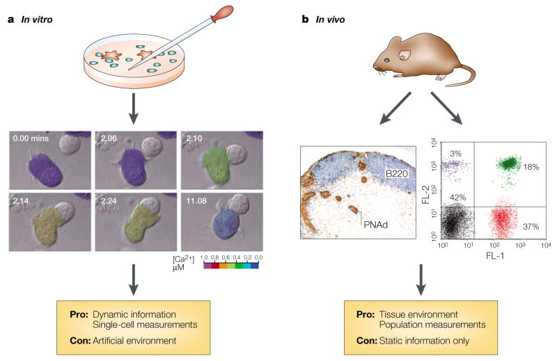
a | In vitro-imaging approaches allow the visualization of cellular and molecular details — illustrated here by the example of an antigen-presenting cell triggering a calcium signal in an interacting T cell. b | Immunohistological and cytometric in vivo methods have made possible the study of cell populations at discrete time points; however, these techniques do not allow the visualization of the dynamic behaviour of single living cells in their native environment. Images reproduced a | from REF. , with permission from Elsevier Science © (1996), and b | by personal communication from J. Cyster and T. Okada.
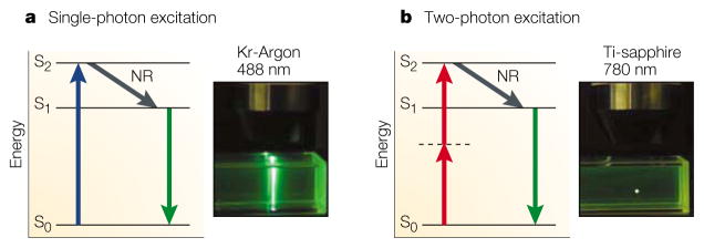
a | Single-photon excitation. Individual photons of high-energy blue light (wavelength, λ = 488 nm) from a krypton-argon (Kr-Argon) laser excite fluorophores in the sample. After an electron in the fluorophore jumps from the energy ground state (S0) to the excited state (S2) (blue arrow), it loses energy rapidly owing to non-radiative relaxation (NR). Subsequently, fluorescence emission occurs at a longer wavelength than the excitation light (Stokes shift) as the electron falls back to the ground state (green arrow). Because excitation involves single photons, fluorescence is emitted along the whole path of a laser beam focused on a sample of fluorescent dye (inset photograph). b | Two-photon excitation. Two infrared photons (λ = 780 nm) from a pulsed titanium-sapphire (Ti-sapphire) laser are absorbed simultaneously (red arrows) to excite the fluorophore; light is emitted in the same manner as for single-photon excitation (green arrow). However, because of the quadratic relationship between excitation intensity and fluorescence emission, light is emitted only at the focal point of the focused laser beam (inset photograph).
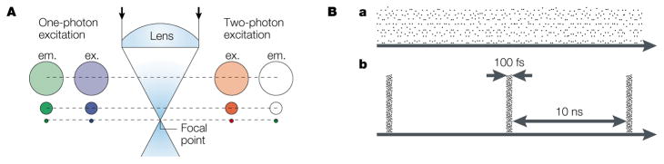
A | Spatial compression of photons by an objective lens restricts two-photon excitation to the focal spot. The diagram compares one-photon excitation (with blue light) with two-photon excitation (with infrared light). In both cases, the excitation intensity (ex.), which corresponds to the density of photons, is greatest at the focal spot and declines away from this point as the light subtends circles of progressively increasing diameter. For one-photon excitation, green-fluorescence emission (em.) occurs everywhere along the path of the beam, with an intensity that is linearly proportional to that of the excitation light. By contrast, two-photon excitation evokes fluorescence at the focal spot only. The rest of the sample is exposed only to low-energy infrared photons, which fail to excite the fluorophore and can pass through biological tissue with minimal scattering. B | Temporal compression of photons into short packets during femtosecond pulses achieves the high photon densities that are required for two-photon excitation and prevents the sample from being destroyed. In comparison to a continuous laser beam (a), a pulsed laser (b) of the same average power (mean number of photons per second) concentrates photons into brief bursts of much greater instantaneous power. For a mode-locked titanium-sapphire laser, each pulse lasts ~100 femtoseconds (fs), with a gap of 10 nanoseconds (ns) between pulses. So, the temporal compression is by a factor of 105 and, because of the quadratic relationship for two-photon excitation, fluorescence is enhanced by a factor of 1010.
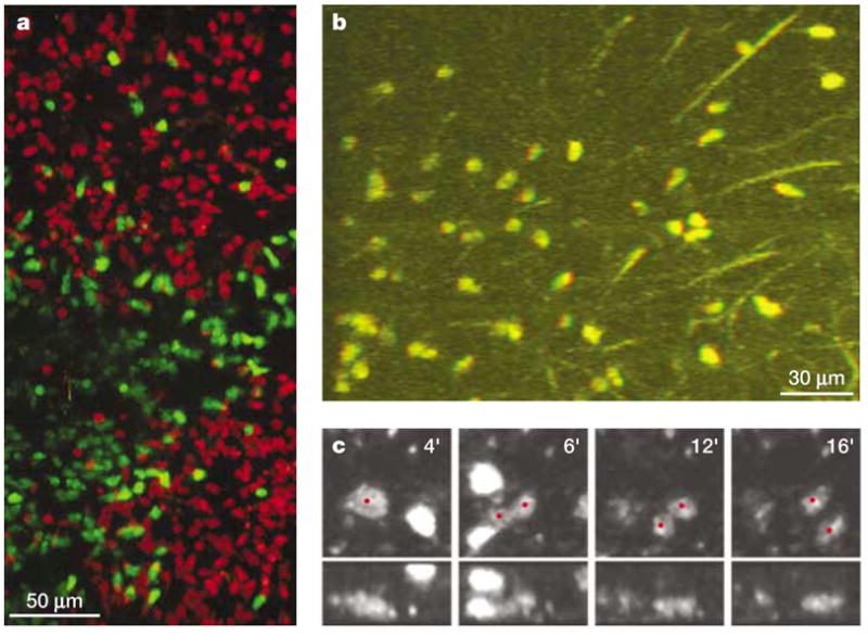
Multi-dimensional imaging data can provide crucial information for understanding the complex behaviour of cells in tissue environments, but they create challenges for analysis and representation in print media. a | Maximum-intensity projection of a 200-μm deep section of an intact lymph node, showing two follicles and an interfollicular space, which contain B cells (red) and T cells (green), respectively. b | A single frame taken from a stereo movie of T cells crawling in a meshwork of reticular fibres (see http://crt.biomol.uci.edu/TCellMovie/stereofiber.mov ) that conveys depth information when viewed with red/green stereo glasses. 51-μm thick section. c | An in vivo proliferation event inside the lymph node is verified by inspecting the time course of the event (left to right, in minutes) in two different reconstructions (top and bottom, 0° and 90° rotations of the z-axis). At 4 minutes, the red dot indicates a rounded, enlarged cell just before division. At 5–6 minutes, a furrow develops in the middle of the non-motile dividing cell, followed by complete separation into half-sized cells and full development of motility by 12–16 minutes (see http://crt.biomol.uci.edu/TCellMovie/dividing.mov ).
References
-
- Donnadieu E, Bismuth G, Trautmann A. Antigen recognition by helper T cells elicits a sequence of distinct changes of their shape and intracellular calcium. Curr Biol. 1994;4:584–595. - PubMed
-
- Negulescu PA, Krasieva TB, Khan A, Kerschbaum HH, Cahalan MD. Polarity of T-cell shape, motility and sensitivity to antigen. Immunity. 1996;4:421–430. This study describes the T cell as a motile, polarized antigen sensor, for which Ca2+ functions as a stop signal. - PubMed
Publication types
MeSH terms
Grants and funding
LinkOut - more resources
Full Text Sources
Other Literature Sources
Medical
