. 2002 Nov;110(9):1243-50.
doi: 10.1172/JCI15736.

Selective parasympathetic innervation of subcutaneous and intra-abdominal fat--functional implications

Affiliations

Selective parasympathetic innervation of subcutaneous and intra-abdominal fat--functional implications

Felix Kreier et al. J Clin Invest. 2002 Nov.

Abstract

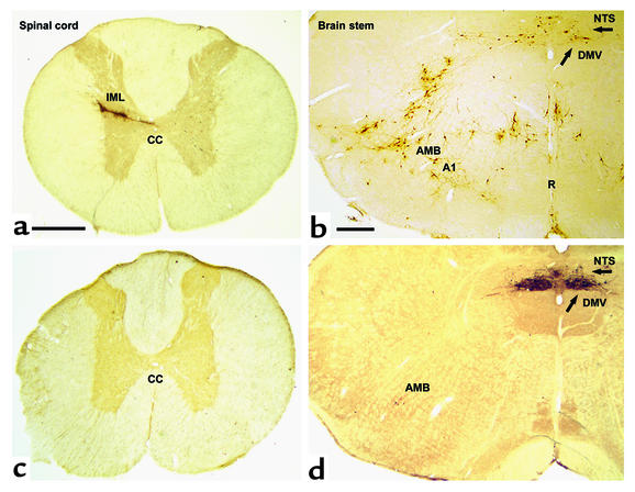
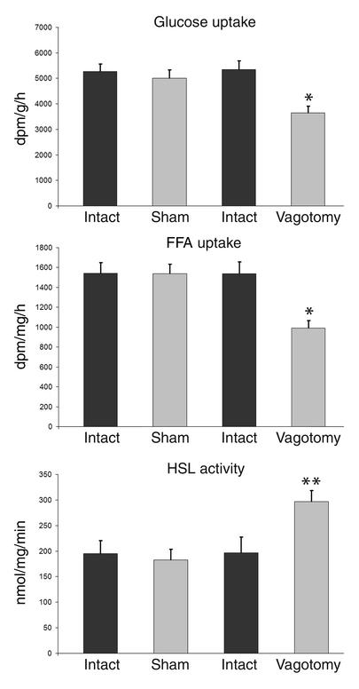
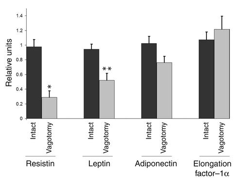
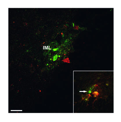
The wealth of clinical epidemiological data on the association between intra-abdominal fat accumulation and morbidity sharply contrasts with the paucity of knowledge about the determinants of fat distribution, which cannot be explained merely in terms of humoral factors. If it comes to neuronal control, until now, adipose tissue was reported to be innervated by the sympathetic nervous system only, known for its catabolic effect. We hypothesized the presence of a parasympathetic input stimulating anabolic processes in adipose tissue. Intra-abdominal fat pads in rats were first sympathetically denervated and then injected with the retrograde transneuronal tracer pseudorabies virus (PRV). The resulting labeling of PRV in the vagal motor nuclei of the brain stem reveals that adipose tissue receives vagal input. Next, we assessed the physiological impact of these findings by combining a fat pad-specific vagotomy with a hyperinsulinemic euglycemic clamp and RT-PCR analysis. Insulin-mediated glucose and FFA uptake were reduced by 33% and 36%, respectively, whereas the activity of the catabolic enzyme hormone-sensitive lipase increased by 51%. Moreover, expression of resistin and leptin mRNA decreased, whereas adiponectin mRNA did not change. All these data indicate an anabolic role for the vagal input to adipose tissue. Finally, we demonstrate somatotopy within the central part of the autonomic nervous system, as intra-abdominal and subcutaneous fat pads appeared to be innervated by separate sympathetic and parasympathetic motor neurons. In conclusion, parasympathetic input to adipose tissue clearly modulates its insulin sensitivity and glucose and FFA metabolism in an anabolic way. The implications of these findings for the (patho)physiology of fat distribution are discussed.


Figures

Figure 1
Figure 1
Transverse section of the spinal cord (at Th7) and the rat brain stem at the level of the obex shows spinal cord and brain stem neurons projecting into adipose tissue. Transneuronal retrograde tracing by PRV injection into retroperitoneal fat in rats before (a and b) and after (c and d) sympathetic denervation of adipose tissue. In a and b (PRV tracing from adipose tissue before denervation), since both sympathetic and parasympathetic fibers are intact, PRV is seen to spread via the vagus and the sympathetic nerves. Interestingly, the route via the IML is favored in intact animals such that second-order neurons in the brain stem are already evident when the first-order parasympathetic motor neurons appear in the DMV (arrow). In b, the A1 region, the raphe nucleus (R), and the nucleus of the solitary tract (NTS) project into the sympathetic motor neurons. In c (with d, showing PRV tracing after sympathetic denervation of the left retroperitoneal fat pad), there is no labeling of PRV in the IML. In the brain stem shown in d, neurons are clearly visible in the parasympathetic motor nuclei: DMV and caudal part of the AMB. CC, central canal. Bar in a and c = 0.5 mm. Bar in b and d = 0.4 mm.
Figure 2
Figure 2
Uptake of glucose and FFA, and HSL activity in adipose tissue after parasympathetic denervation. The left retroperitoneal fat pad was either parasympathetically denervated (n = 6) or sham operated (n = 6). Using a hyperinsulinemic euglycemic clamp, the uptake of 3H-2-deoxy-D-glucose and 14C-palmitate and the activity of the catabolic enzyme HSL were defined. Under these hyperinsulinemic conditions, glucose uptake in the denervated fat pad was reduced by 33% (by Mann-Whitney U test, *P = 0.02) and FFA by 36% (Mann-Whitney U test, *P = 0.02); HSL activity increased by 51% (Mann-Whitney U test, **P = 0.03). Thus, parasympathetic denervation of adipose tissue shifts the metabolism to a catabolic state: uptake of substrate is decreased, while lipolysis increases. Values are expressed as mean ± SEM. dpm, disintegrations per minute.
Figure 3
Figure 3
Hormone mRNA expression in adipose tissue after parasympathetic denervation. The left retroperitoneal fat pad was parasympathetically denervated (n = 9) and compared with the right intact pad for the expression of mRNA of resistin, leptin, adiponectin, and elongation factor–1α (as a reference gene) by means of real-time RT-PCR. Sham-operated animals were used as control (n = 5). While resistin and leptin mRNA expression was reduced (–71%, Mann-Whitney U test, *P = 0.001; –45%, Mann-Whitney U test, **P = 0.004, respectively), adiponectin and reference mRNA did not change significantly. Thus, parasympathetic denervation of adipose tissue specifically changes mRNA expression of fat-derived hormones. One relative unit is the equivalent cDNA corresponding with 0.1 μg per well of the pooled cDNA of the control fat pads. Values are expressed as mean ± SEM.
Figure 4
Figure 4
Somatotopic organization of the parasympathetic nervous system. Laser scanning photomicrograph of transverse sections of the brain stem. The central canal is on the right side. Vagal motor neurons project into one fat compartment only (subcutaneous or intra-abdominal). PRV (stained green) was injected into the intra-abdominal fat compartment after sympathetic denervation. At the same time, FluoroGold (stained red) was injected into the subcutaneous fat compartment. Both tracers were transported back to the dorsal motor nucleus DMV and AMB in different neuron populations. Somatotopic segregation can be observed within the DMV. Bar = 50 μm.
Figure 5
Figure 5
Somatotopic organization of the sympathetic nervous system. Sympathetic motor neurons project into one fat compartment only (subcutaneous or intra-abdominal). Two strains of PRV were injected simultaneously into the intra-abdominal fat compartment (mesenterial fat) and the subcutaneous fat compartment (subcutaneous inguinal fat). Confocal laser scanning photomicrograph of transverse thoracic spinal cord sections (Th5–Th10). Both tracers were transported back to the IML and show clear separation of the different tracers (red/green). Insert (control): Injection of both tracers into the same mesenterial fat pad resulted in colocalization of the two tracers (yellow). Specific laser analysis of the indicated neuron (arrow) also showed colocalization of the tracers with a strong signal of FITC (green) and a much weaker signal of CY3 (red). Bar = 50 μm.

Comment in

References

    1. Wajchenberg BL. Subcutaneous and visceral adipose tissue: their relation to the metabolic syndrome. Endocr Rev. 2000;21:697–738. - PubMed
    1. Kalsbeek A, et al. The suprachiasmatic nucleus generates the diurnal changes in plasma leptin levels. Endocrinology. 2001;142:2677–2685. - PubMed
    1. Ludwig DS, et al. Melanin-concentrating hormone overexpression in transgenic mice leads to obesity and insulin resistance. J Clin Invest. 2001;107:379–386. - PMC - PubMed
    1. Buijs RM, Kalsbeek A. Hypothalamic integration of central and peripheral clocks. Nat Rev Neurosci. 2001;2:521–526. - PubMed
    1. Bray GA, York DA. Hypothalamic and genetic obesity in experimental animals: an autonomic and endocrine hypothesis. Physiol Rev. 1979;59:719–809. - PubMed