. 2002 Nov;110(9):1309-18.
doi: 10.1172/JCI15451.

Transport of paclitaxel (Taxol) across the blood-brain barrier in vitro and in vivo

Affiliations

Transport of paclitaxel (Taxol) across the blood-brain barrier in vitro and in vivo

Stephan Fellner et al. J Clin Invest. 2002 Nov.

Abstract

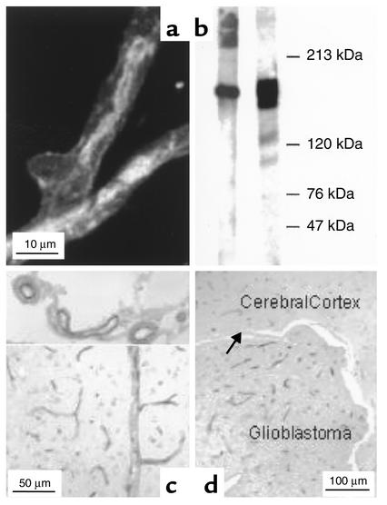
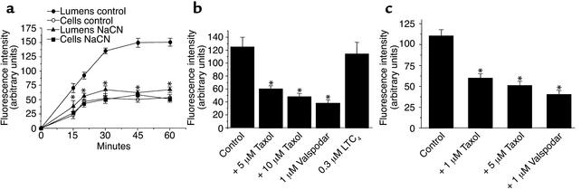
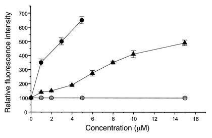
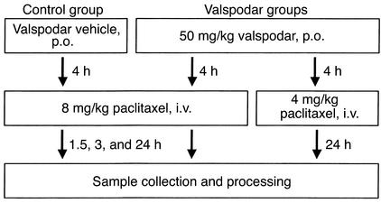
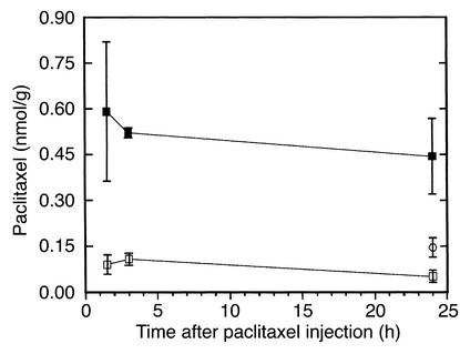
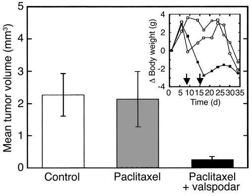
Paclitaxel concentrations in the brain are very low after intravenous injection. Since paclitaxel is excluded from some tumors by p-glycoprotein (p-gp), the same mechanism may prevent entry into the brain. In vitro, paclitaxel transport was examined in capillaries from rat brains by confocal microscopy using BODIPY Fl-paclitaxel. Western blots and immunostaining demonstrated apical expression of p-gp in isolated endothelial cells, vessels, and tissue. Secretion of BODIPY Fl-paclitaxel into capillary lumens was specific and energy-dependent. Steady state luminal fluorescence significantly exceeded cellular fluorescence and was reduced by NaCN, paclitaxel, and SDZ PSC-833 (valspodar), a p-gp blocker. Leukotriene C(4) (LTC(4)), an Mrp2-substrate, had no effect. Luminal accumulation of NBDL-cyclosporin, a p-gp substrate, was inhibited by paclitaxel. In vivo, paclitaxel levels in the brain, liver, kidney, and plasma of nude mice were determined after intravenous injection. Co-administration of valspodar led to increased paclitaxel levels in brains compared to monotherapy. Therapeutic relevance was proven for nude mice with implanted intracerebral human U-118 MG glioblastoma. Whereas paclitaxel did not affect tumor volume, co-administration of paclitaxel (intravenous) and PSC833 (peroral) reduced tumor volume by 90%. Thus, p-gp is an important obstacle preventing paclitaxel entry into the brain, and inhibition of this transporter allows the drug to reach sensitive tumors within the CNS.


Figures

Figure 1
Figure 1
(a) Immunostaining of p-gp in a freshly isolated rat brain capillary (primary Ab: MAB C219; secondary Ab: FITC-conjugated rabbit anti-mouse IgG). (b) P-gp detection by Western blot using the MAB C219. Left lane: brain capillaries; right lane: p-gp overexpressing cell line (MDR+)P388 as positive control. (c) Immunostaining of coronar sections of the brains of nude mice, using the polyclonal AB-1 Ab. Healthy animal, with P-gp expression in capillaries and larger blood vessels in the cerebrum (bottom) and the submeningeal arteries and veins (top). (d) Intracerebral human glioblastoma (U-87 MG) with immunopositive capillary endothelia and negative tumor cells. The arrow points to the ependyma.
Figure 2
Figure 2
(a) Time-dependent accumulation of BODIPY Fl-paclitaxel in cells and lumens of isolated rat brain capillaries in the absence and presence of 1 mM NaCN. Means ± SEM, n = 10–12, P = 0.05. (b) Accumulation of BODIPY Fl-paclitaxel and NBDL-CS (c), respectively, in the lumens of isolated capillaries in the absence and presence of unlabeled paclitaxel, the p-gp blocker valspodar, and the mrp2 substrate LTC4. The fluorescent compounds were added at concentrations of 1 μM. Means ± SEM, n = 10–12. *Significantly different from controls; P = 0.05.
Figure 3
Figure 3
Increase of intracellular fluorescence intensity (free calcein) in monolayers of porcine brain capillary endothelial cells. Presence of the p-gp substrates valspodar (filled circles) and paclitaxel (filled triangles) results in an increased fluorescence intensity, whereas penicillin (open circles) has no effect (means ± SEM, n = 6).
Figure 4
Figure 4
Application scheme of PSC 833 and paclitaxel to nude mice. Each group consisted of three to four nude mice per observation time after intravenous injection.
Figure 5
Figure 5
Effect of valspodar coadministration on the paclitaxel brain levels in nude mice after intravenous injection of 8 mg/kg paclitaxel. Four hours before intravenous application the animals were treated with the vehicle of valspodar by mouth (control, open squares) or 50 mg/kg valspodar by mouth (valspodar group, filled squares). Brain levels for a valspodar group with reduced paclitaxel dose (4 mg/kg, open circles) were investigated 24 hours after intravenous injection. Means ± SEM, n = 3–4. Compared with the respective controls, all values in the valspodar groups were significantly (P ≤ 0.05) higher.
Figure 6
Figure 6
Effect of valspodar/paclitaxel coadministration on the intracerebral growth of the human U 118 MG glioblastoma in nude mice. Controls received no medication, the paclitaxel group received the vehicle of valspodar 4 hours before injection of paclitaxel, and the paclitaxel-valspodar group received 50 mg/kg valspodar by mouth before injection of paclitaxel. Animals were treated in this way on day 8 (3 mg/kg paclitaxel) and day 15 (2 mg/kg paclitaxel) after implantation of tumors. Thirty-five days after tumor implantation the brains of the animals were collected and the tumor volumes determined morphometrically. The figure shows mean values ± SEM (n = 5). Inset: Change of mean body weight as an indicator of toxicity. Control, open squares; paclitaxel, open circles; paclitaxel-valspodar, filled squares. The arrows indicate drug administration.

References

    1. Kroger N, Achterrath W, Hegewisch-Becker S, Mross K, Zander AR. Current options in treatment of anthracycline-resistant breast cancer. Cancer Treat Rev. 1999;25:279–291. - PubMed
    1. Nathan FE, Berd D, Sato T, Mastrangelo MJ. Paclitaxel and tamoxifen: an active regimen for patients with metastatic melanoma. Cancer. 2000;88:79–87. - PubMed
    1. Muggia FM, et al. Phase III randomized study of cisplatin versus paclitaxel versus cisplatin and paclitaxel in patients with suboptimal stage III or IV ovarian cancer: a gynecologic oncology group study. J Clin Oncol. 2000;18:106–115. - PubMed
    1. Papadimitriou CA. Paclitaxel, cisplatin, and epirubicin first-line chemotherapy in stage III and IV ovarian carcinoma: long-term results of a phase II study. Cancer. 2000;89:1547–1554. - PubMed
    1. Belani CP. Paclitaxel and docetaxel combinations in non-small cell lung cancer. Chest. 2000;117(Suppl 1):144S–151S. - PubMed

Publication types