. 2002 Nov;110(9):1349-58.
doi: 10.1172/JCI15614.

P311 induces a TGF-beta1-independent, nonfibrogenic myofibroblast phenotype

Affiliations

P311 induces a TGF-beta1-independent, nonfibrogenic myofibroblast phenotype

Desi Pan et al. J Clin Invest. 2002 Nov.

Abstract

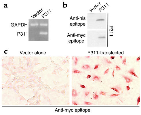
P311, also called PTZ17, was identified by suppressive subtraction hybridization as potentially involved in smooth muscle (SM) myogenesis. P311 is an 8-kDa protein with several PEST-like motifs found in neurons and muscle. P311 transfection into two fibroblast cell lines, NIH 3T3 and C3H10 T1/2, induced phenotypic changes consistent with myofibroblast transformation, including upregulation of SM alpha-actin and SM22, induction of FGF-2, VEGF, PDGF, and PDGF receptors, upregulation of integrins alpha3 and alpha5, and increased proliferation rate. The P311-mediated changes differed, however, from the well-characterized myofibroblast in that P311 inhibited TGF-beta1, TGF-beta receptor 2, and TGF-beta1-activating MMP-2 and MMP-9, with the resultant decrease in collagen 1 and 3 expression. The effect of P311 on collagen was overcome by exogenous TGF-beta1, indicating that the cells were responsive to TGF-beta1 paracrine stimulus. In support of a role for P311 in vivo, immunohistochemical examination of human wounds showed P311 only in myofibroblasts and their activated precursors. To our knowledge, these studies are the first to implicate P311 in myofibroblast transformation, to demonstrate that transformation may occur independently of TGF-beta1, and to suggest that P311 may prevent fibrosis.


Figures

Figure 1
Figure 1
Transfection of myc-his–tagged P311 into NIH-3T3 cells. (a and b) RT-PCR and Western blot showing no P311 expression in cells transfected with vector alone and synthesis of P311 mRNA and protein upon P311 transfection. (c) Immunostaining with anti-tagging epitope showing that NIH-3T3 cells transfected with vector alone are negative (left panel), whereas P311-transfected cells (right panel) show rather uniform synthesis of P311 after a month of G418 treatment for selection of transfected cells.
Figure 2
Figure 2
Transfection of P311 into NIH-3T3 cells stimulates muscle-specific transcription factors. (a) RT-PCR showing mRNA for muscle-related transcription factors in cells transfected with empty vector or transfected with P311. Cells expressing P311 show higher levels of muscle transcription factors, whether related to striated or SM differentiation, except for Myf5, which was downregulated. (b) NIH 3T3 cells transiently transfected with P311 and double immunostained for P311 (immunoperoxidase, brown in the upper panel) and various myogenic transcription factors (green immunofluorescence in lower panel). Notice upregulation of muscle transcription factors in the nucleus of P311-transfected cells (white arrows). (c) Immunoprecipitation (P311) and immunoblots (others) demonstrating upregulation of muscle transcription factors by transfected P311 compared with control cells transfected with empty vector. The same membrane used for detection of SRF and reprobing for MEF2 was stained blue with Coomassie to show protein loading. Mgnin, myogenin.
Figure 3
Figure 3
Transfection of P311 into NIH 3T3 cells stimulates expression of SM α-actin and SM22. (a) RT-PCR showing mRNA for three SM-specific markers in cells transfected with empty vector or transfected with P311. Notice that P311 upregulated SM α-actin message, induced SM22 message and did not alter significantly the mRNA for SM-myosin, present in both control and P311-expressing cells. (b) Cells expressing P311 do not show either STM-specific markers such as STM α-actin and myoglobin or desmin, a protein present in both STM and SM. (c) Immunoblot demonstrating upregulation of SM α-actin and induction of SM22 in P311-transfected cells and absence of other SM and STM markers. The membrane used for SM22 detection was stained with Coomassie blue to show equal protein loading.
Figure 4
Figure 4
Transfection of P311 into C310 T1/2 cells stimulates muscle-specific transcription factors and induces SM α-actin synthesis. (a) RT-PCR showing mRNA for P311 and muscle-specific transcription factors in cells transfected with empty vector or transfected with P311. P311-expressing cells have higher levels of muscle-specific transcription factors than their control counterparts, with the exception of Myf5, which is downregulated. (b) Immunoblots showing increase in the synthesis of muscle-related transcription factors in P311-transfected cells compared with controls. The membrane used for detection of MyoD was stained with Coomassie blue to show equal protein loading. (c) Immunoblots demonstrating P311-induced upregulation of SM α-actin but absence of other SM and STM markers. (d) RT-PCR showing presence of mRNA for desmin and SM-myosin in control and P311-expressing cells, with some increment in SM-myosin message, which was consistent among experiments.
Figure 5
Figure 5
P311 induces cellular changes in NIH 3T3 cells characteristic of myofibroblast transformation. (ac) RT-PCR and microarray analysis demonstrating stimulation of VEGF, PDGF, and FGF-2 (a), PDGF receptors α (PDGF-Rα) and β (PDGF-Rβ) (b), and integrins α3 (Int α3) and α5 (Int α5) (c) in P311-transfected cells compared with controls transfected with vector alone. Integrin α2 (Int α2) was not expressed in either of them. There were two different cDNA microarray membranes; therefore the GAPDH internal control for each was included in the figure. (d) Histogram showing the increase in each respective mRNA in P311-expressing cells as established by densitometry of microarray dots. (e) Histogram showing increased proliferation rate for P311-expressing cells when cultured in the presence of serum and better survival in serumless conditions.
Figure 6
Figure 6
P311 inhibits TGF-β1 and TGF-βR2 in NIH 3T3 and C3H10 T1/2 cells and decreases collagen 1 and 3 expression. (a) RT-PCR and immunoprecipitation followed by immunoblotting (lower panels) showing downregulation of TGF-β1 in P311-expressing cells compared with controls. (b) RT-PCR and Western blotting demonstrating P311-induced downregulation of TGF-βR2 but not of TGF-βR1 in NIH 3T3 (upper panels) and C3H10 T1/2 (lower panels). Untreated (untreat) cells were used as an additional control for C3H10 T1/2 cells in the lower panels. (c) RT-PCR showing significant collagen 1 (CN1) and 3 (CN3) downregulation in P311-expressing cells (upper panels) and inhibition of MMP-2 and MMP-9, which participate in TGF-β1 activation and inhibition of MMP-1 that is upregulated by TGF-β1. (d) RT-PCR performed on control and P311-expressing NIH 3T3 cells after 48 hours in culture with or without TGF-β1 treatment demonstrating reversal of P311-induced downregulation in collagen expression by exogenous TGF-β1.
Figure 7
Figure 7
P311 is expressed in human visceral and vascular SM cells and myofibroblasts. All the sections have been double immunostained for P311 and SM α-actin, the latter to identify SM and myofibroblasts. (a) P311 is present in both visceral (intestine) and vascular SM (first and second panels down, respectively). P311 is not synthesized by fibroblasts (third panels), but is produced by myofibroblasts (fourth panels). (b) Immunohistochemical studies performed at an early stage of wound repair demonstrate P311 in most of the activated fibroblasts, also referred to as premyofibroblasts (SM α-actin–negative myofibroblast precursors, arrow in upper left photo). In more advanced stages of wound healing (two middle panels), P311 is detected in myofibroblasts (activated, SM α-actin–positive cells). Arrowhead in the second left photo points to a regular fibroblast present in the same field. Notice its smaller size, lack of discernible nucleoli, and negativity for P311. Once myofibroblasts disappear and the wound contains only regular fibroblasts, P311 is no longer detected (lowest panels).

References

    1. Relan NK, Yang Y, Beqaj S, Miner JH, Schuger L. Cell elongation induces laminin alpha2 chain expression in mouse embryonic mesenchymal cells. Role in visceral myogenesis. J Cell Biol. 1999;147:1341–1350. - PMC - PubMed
    1. Yang Y, Relan NK, Przywara DA, Schuger L. Embryonic mesenchymal cells share the potential for smooth muscle differentiation: myogenesis is controlled by the cell’s shape. Development. 1999;126:3027–3033. - PubMed
    1. Yang Y, Beqaj S, Kemp P, Ariel I, Schuger L. Stretch-induced alternative splicing of serum response factor promotes bronchial myogenesis and is defective in lung hypoplasia. J Clin Invest. 2000;106:1321–1330. - PMC - PubMed
    1. Liu J, Beqaj S, Yang Y, Honore B, Schuger L. Heterogeneous nuclear ribonucleoprotein-H plays a suppressive role in visceral myogenesis. Mech Dev. 2001;104:79–87. - PubMed
    1. Studler JM, Glowinski J, Levi-Strauss M. An abundant mRNA of the embryonic brain persists at a high level in cerebellum, hippocampus and olfactory bulb during adulthood. Eur J Neurosci. 1993;5:614–623. - PubMed

Publication types