. 2002 Nov 4;196(9):1175-88.
doi: 10.1084/jem.20020735.

Failure to censor forbidden clones of CD4 T cells in autoimmune diabetes

Affiliations

Failure to censor forbidden clones of CD4 T cells in autoimmune diabetes

Sylvie Lesage et al. J Exp Med. .

Abstract

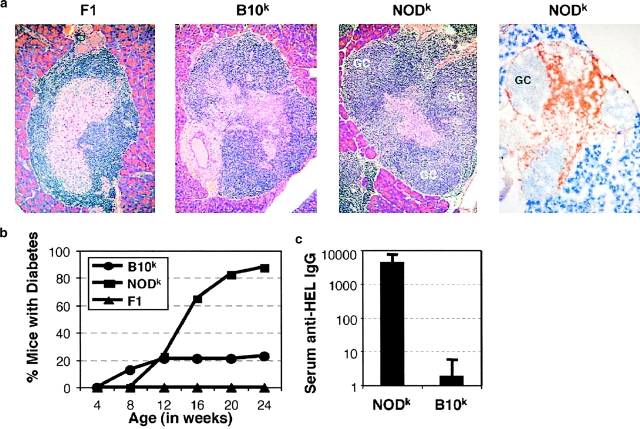
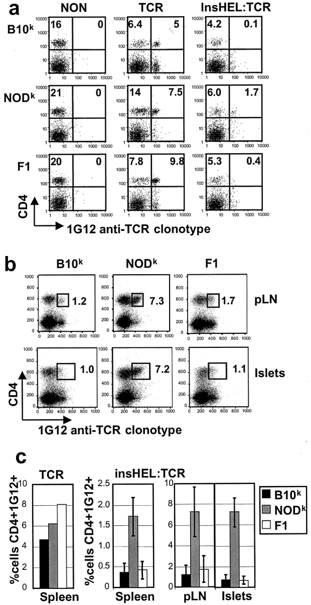
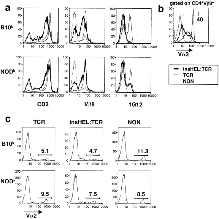
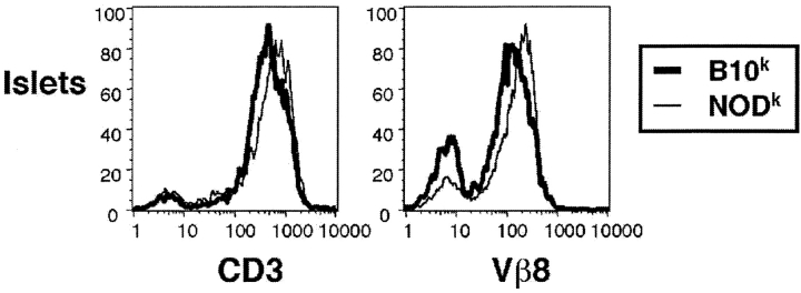
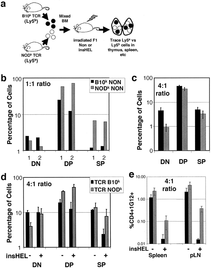
Type 1 diabetes and other organ-specific autoimmune diseases often cluster together in human families and in congenic strains of NOD (nonobese diabetic) mice, but the inherited immunoregulatory defects responsible for these diseases are unknown. Here we track the fate of high avidity CD4 T cells recognizing a self-antigen expressed in pancreatic islet beta cells using a transgenic mouse model. T cells of identical specificity, recognizing a dominant peptide from the same islet antigen and major histocompatibility complex (MHC)-presenting molecule, were followed on autoimmune susceptible and resistant genetic backgrounds. We show that non-MHC genes from the NOD strain cause a failure to delete these high avidity autoreactive T cells during their development in the thymus, with subsequent spontaneous breakdown of CD4 cell tolerance to the islet antigen, formation of intra-islet germinal centers, and high titre immunoglobulin G1 autoantibody production. In mixed bone marrow chimeric animals, defective thymic deletion was intrinsic to T cells carrying diabetes susceptibility genes. These results demonstrate a primary failure to censor forbidden clones of self-reactive T cells in inherited susceptibility to organ-specific autoimmune disease, and highlight the importance of thymic mechanisms of tolerance in organ-specific tolerance.


Figures

Figure 1.
Figure 1.
Islet-reactive T cells cause germinal center formation, autoantibodies, and diabetes in NODk mice. (a) Pancreatic islets stained with H&E from TCR/InsHEL mice of the indicated genotypes. Right panel is a frozen section stained with peanut agglutinin (blue) and anti-IgD (brown). GC, germinal center. (b) Onset of diabetes in TCR/insHEL female mice of the indicated genotypes. Data represent >20 mice per group. (c) HEL-binding IgG titre in serum from diabetic double transgenic mice, collected within 2 wk of diabetes onset. Titres are expressed in arbitrary units relative to a reference immune sera from HEL-immunized nontransgenic mice set at 100 units.
Figure 2.
Figure 2.
Failure to eliminate islet-reactive CD4 T cells in NODk mice. (a) Spleen cells from 6–12-wk old nondiabetic female mice of the indicated genotypes were stained with 1G12 anti-TCR clonotype and antibody to CD4. The percentage of lymphocytes falling within the quadrants is shown. (b) The same analysis performed on cells from pancreatic lymph nodes (pLN) and pancreatic islets of 6–12-wk old nondiabetic TCR/insHEL mice. (c) Data collected as above compiled from multiple animals of the indicated genotypes.
Figure 3.
Figure 3.
Failure to downregulate TCR expression in splenocytes of NODk animals. (a) Spleen cells from nondiabetic female mice of various genotypes were stained with antibodies to CD4, CD3, Vβ8, and 1G12 clonotype. Histograms show profiles of CD4-positive cells from mice of the indicated genotypes. (b) Vα3 expression on CD4+Vβ8+ spleen cells from B10k mice of the indicated genotypes. (c) Staining for an endogenous TCR-α chain, Vα2, on CD4+ spleen cells. The percentage of CD4+ cells that are Vα2+ is indicated.
Figure 4.
Figure 4.
High TCR levels on lymphocytes in the islets of NODk mice. Islets from nondiabetic female mice were purified and analyzed as in Fig. 3 a.
Figure 5.
Figure 5.
T cell activation in the islets is specific to CD4+clonotype+ NODk T cells. Lymphocytes from pancreatic lymph nodes and islets of two nondiabetic TCR/insHEL female mice of B10k or NODk background were stained with CD4, 1G12 clonotype, and the activation marker CD69. The percentage of lymphocytes falling within the quadrants is indicated. Histograms show profiles of CD69 expression on clonotype-positive or clonotype-negative CD4 cells, overlaying cells from the islet and pancreatic lymph node of the same animal.
Figure 6.
Figure 6.
Failure to delete islet-reactive T cells in the thymus of NODk animals. (a) Thymocytes from 6–12-wk old nondiabetic female mice of the indicated genotypes were stained with 1G12 anti-TCR clonotype, and antibodies to CD4 and CD8. The percentage of CD4+CD8 and CD4+CD8+ thymocytes are shown. (b) The 1G12 profiles are shown for CD4+CD8 mature thymocytes. 1G12 staining cannot be detected on CD4+CD8+ thymocytes. (c) Absolute number of CD4+CD8+ thymocytes was calculated for each group. From left to right, there were exactly 4, 6, 16, 3, 5, 13, and 10 mice, respectively. (d) Compilation of the number of CD4+1G12+ thymocytes in each group.
Figure 7.
Figure 7.
Failure to downregulate TCR expression in CD4+ thymocytes of NODk animals. (a) Thymocytes from nondiabetic female mice of various genotypes were stained with antibodies to CD4, CD8, and 1G12 clonotype, together with either CD3 or Vβ8. All histograms show profiles of CD4+CD8 cells, except for the graphs on the right which are gated on CD4+CD8+ (DP) thymocytes.
Figure 8.
Figure 8.
Increased CD4+1G12+ thymocytes in NODk insHEL/TCR mice is not due to mature T cell recirculation to the thymus. (a) Absolute number of CD4+1G12+ mature thymocytes in insHEL/TCR double transgenic on B10k, NODk, and F1 background plotted versus age. (b) Thymocytes from insHEL/TCR double transgenic mice were stained with 1G12, CD4, CD8, and CD24. CD24 profiles are shown for 1G12-positive or -negative CD4+CD8 mature thymocytes.
Figure 9.
Figure 9.
Failure to delete islet-reactive cells in NODk is T cell intrinsic. (a) Design of bone marrow chimera experiment. (b) Nontransgenic Ly5ab recipients were reconstituted with a 1:1 mixture of bone marrow from B10k and NODk nontransgenic mice. The thymocytes were stained with CD4, CD8, Ly5a and Ly5b. Percentage of each thymocyte subsets is shown for two mice. (c) Percentage is as in b, except that the ratio of B10k/NODk bone marrow cells is 4:1. (d) InsHEL-positive or -negative recipients were reconstituted with a 4:1 ratio of B10k/NODk marrow. Percentage of thymocyte subsets is shown from analysis of four nontransgenic and eight insHEL-transgenic recipients. (e) Percentage of peripheral lymphocytes bearing CD4 and the HEL- reactive TCR from each donor genotype from the mice in days.

References

    1. Todd, J.A., and L.S. Wicker. 2001. Genetic protection from the inflammatory disease type 1 diabetes in humans and animal models. Immunity. 15:387–395. - PubMed
    1. Sinha, A.A., M.T. Lopez, and H.O. McDevitt. 1990. Autoimmune diseases: the failure of self tolerance. Science. 248:1380–1388. - PubMed
    1. Bach, J.F., and L. Chatenoud. 2001. Tolerance to islet autoantigens in Type 1 diabetes. Annu. Rev. Immunol. 19:131–161. - PubMed
    1. Adorini, L., S. Gregori, and L.C. Harrison. 2002. Understanding autoimmune diabetes: insights from mouse models. Trends Mol. Med. 8:31–38. - PubMed
    1. Delovitch, T.L., and B. Singh. 1997. The nonobese diabetic mouse as a model of autoimmune diabetes: immune dysregulation gets the NOD. Immunity. 7:727–738. - PubMed

Publication types