

Correlations between visual recognition memory and neocortical and hippocampal glucose metabolism after bilateral rhinal cortex lesions in the baboon: implications for Alzheimer's disease
- PMID: 12417640
- PMCID: PMC6758045
- DOI: 10.1523/JNEUROSCI.22-21-09166.2002
Correlations between visual recognition memory and neocortical and hippocampal glucose metabolism after bilateral rhinal cortex lesions in the baboon: implications for Alzheimer's disease
Erratum in
- J Neurosci. 2002 Dec 15;22(24):1a.. Blaizot A Xavier [corrected to Blaizot Xavier]
Abstract
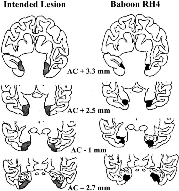
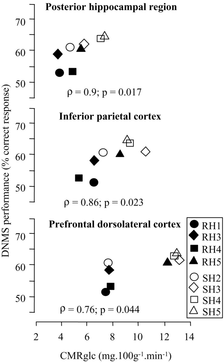
In Alzheimer's disease (AD), the rhinal cortex is the area earliest and most affected by neurofibrillary tangles, and the degree of temporoparietal glucose hypometabolism and rhinal cortex atrophy are both correlated with dementia severity. In monkeys, damage to the rhinal cortex leads to severe impairment in declarative memory, which is also affected preferentially in early AD. To investigate the contribution of rhinal alterations to the interrelationships between cerebral hypometabolism and declarative memory impairment observed in AD, we studied the effects of excitotoxic bilateral rhinal lesions in baboons on cerebral glucose consumption (CMRglc) as measured by positron emission tomography and performance on a visual recognition memory task as assessed in parallel by a delayed nonmatching-to-sample task. We reported previously that these rhinal lesions induce both a long-lasting hypometabolism in several remote brain regions (Meguro et al., 1999) and impaired memory performance (Chavoix et al., 2002). The present analysis indicates that across lesioned and sham baboons, memory scores were significantly positively correlated (p < 0.05; Spearman) with concomitant CMRglc values of several brain areas, such as neocortical associative and posterior hippocampal regions. These findings, reminiscent of those reported in AD, suggest that the neurodegenerative process that affects the rhinal cortex in early AD plays a crucial role in the pattern of brain hypometabolism and consequently in the declarative memory impairments characteristic of this disease.
Figures
References
-
- Aggleton JP, Mishkin M. Visual recognition impairment following medial thalamic lesions in monkeys. Neuropsychologia. 1983;21:189–197. - PubMed
-
- Aigner TG, Mitchell SJ, Aggleton JP, DeLong MR, Struble RG, Price DL, Wenk GL, Pettigrew KD, Mishkin M. Transient impairment of recognition memory following ibotenic-acid lesions of the basal forebrain in macaques. Exp Brain Res. 1991;86:18–26. - PubMed
-
- Blaizot X, Meguro K, Le Mestric C, Constans JM, Luet D, Baron JC, Chavoix C. Combined use of T1-weighted MRI and MRA for stereotaxic lesioning of the nonhuman primate brain: application to the rhinal cortex. Exp Brain Res. 1999;126:31–40. - PubMed
-
- Blaizot X, Landeau B, Baron JC, Chavoix C. Mapping the visual recognition memory network with PET in the behaving baboon. J Cereb Blood Flow Metab. 2000;20:213–219. - PubMed
Publication types
MeSH terms
Substances
LinkOut - more resources
Full Text Sources
Medical