. 2002 Nov 1;22(21):9340-51.
doi: 10.1523/JNEUROSCI.22-21-09340.2002.

Abundant tau filaments and nonapoptotic neurodegeneration in transgenic mice expressing human P301S tau protein

Affiliations

Abundant tau filaments and nonapoptotic neurodegeneration in transgenic mice expressing human P301S tau protein

Bridget Allen et al. J Neurosci. .

Abstract

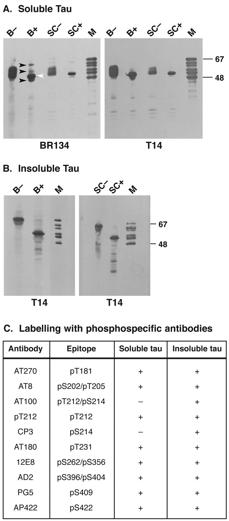
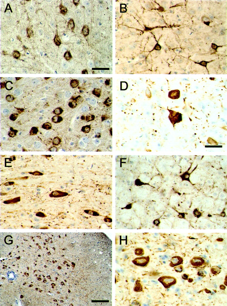
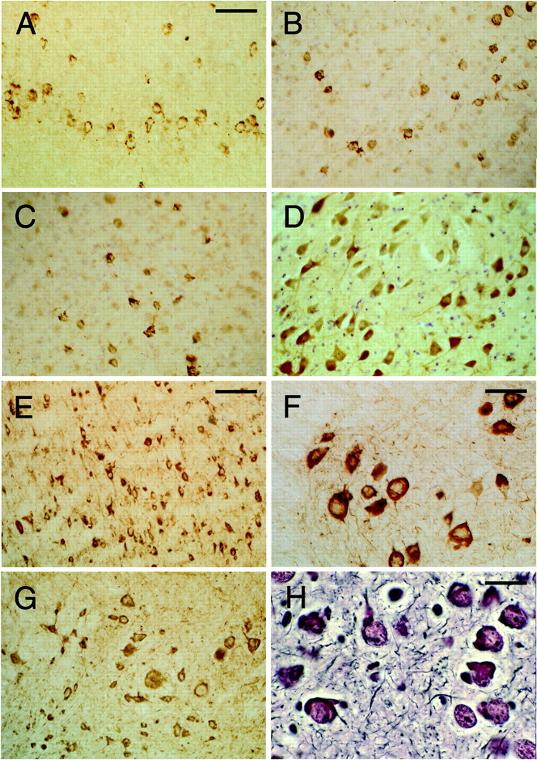
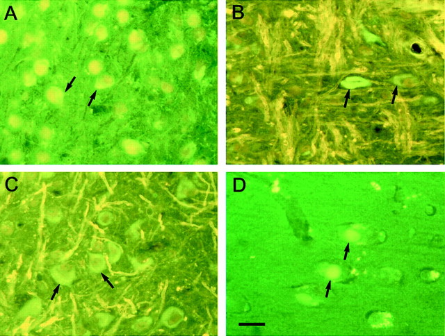
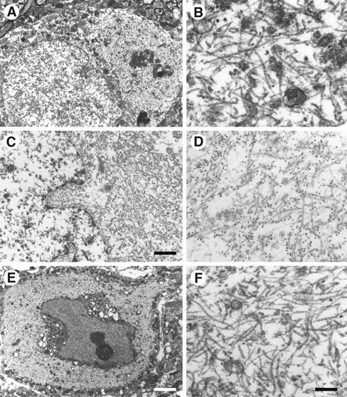
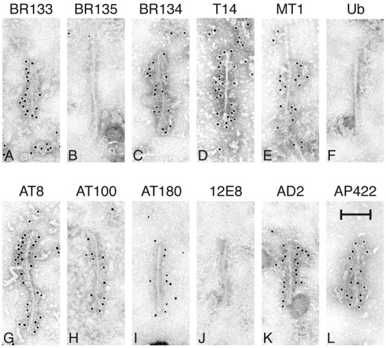
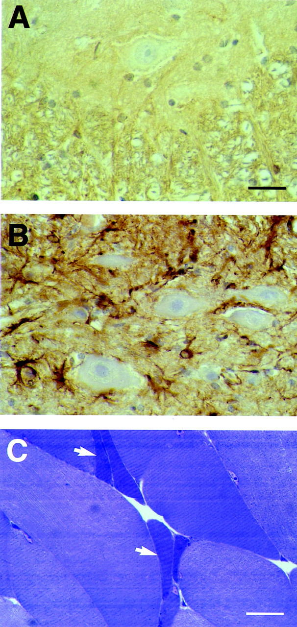
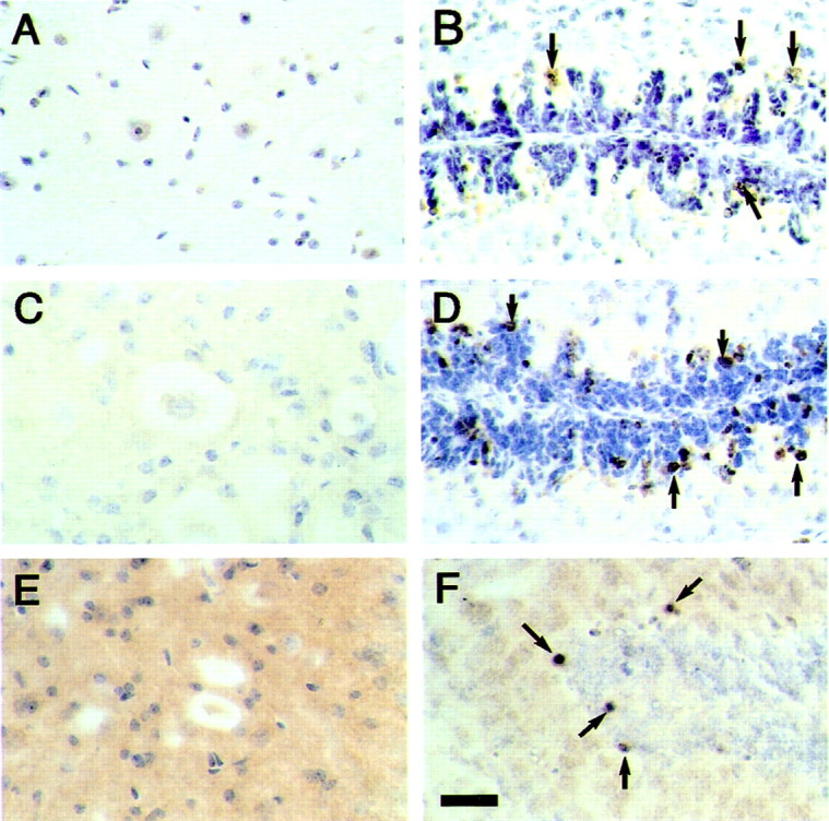
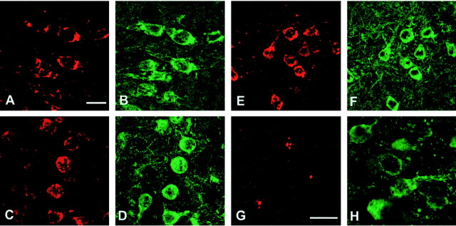
The identification of mutations in the Tau gene in frontotemporal dementia and parkinsonism linked to chromosome 17 (FTDP-17) has made it possible to express human tau protein with pathogenic mutations in transgenic animals. Here we report on the production and characterization of a line of mice transgenic for the 383 aa isoform of human tau with the P301S mutation. At 5-6 months of age, homozygous animals from this line developed a neurological phenotype dominated by a severe paraparesis. According to light microscopy, many nerve cells in brain and spinal cord were strongly immunoreactive for hyperphosphorylated tau. According to electron microscopy, abundant filaments made of hyperphosphorylated tau protein were present. The majority of filaments resembled the half-twisted ribbons described previously in cases of FTDP-17, with a minority of filaments resembling the paired helical filaments of Alzheimer's disease. Sarkosyl-insoluble tau from brains and spinal cords of transgenic mice ran as a hyperphosphorylated 64 kDa band, the same apparent molecular mass as that of the 383 aa tau isoform in the human tauopathies. Perchloric acid-soluble tau was also phosphorylated at many sites, with the notable exception of serine 214. In the spinal cord, neurodegeneration was present, as indicated by a 49% reduction in the number of motor neurons. No evidence for apoptosis was obtained, despite the extensive colocalization of hyperphosphorylated tau protein with activated MAP kinase family members. The latter may be involved in the hyperphosphorylation of tau.


Figures

Fig. 1.
Fig. 1.
Epitopes of anti-tau antibodies. A bar diagram of the 441 aa isoform of human tau is shown, with the microtubule-binding repeat region shown in black. Amino acids recognized by phosphorylation-dependent antibodies are indicated abovethe bar diagram (pS andpT). The epitopes of phosphorylation-independent antibodies are shown below the bar diagram.
Fig. 2.
Fig. 2.
Immunoblot analysis of tau protein in brains and spinal cords from mice of the human P301S tau line. A, Perchloric acid-soluble tau was extracted from the brain (B) and spinal cord (SC) of 5- to 6-month-old mice and immunoblotted with anti-tau antibodies BR134 and T14 before (−) and after (+) alkaline phosphatase treatment.Black arrowheads point to the three mouse tau isoforms, with the white arrowhead pointing to the single human tau isoform. B, Sarkosyl-insoluble tau was extracted from brains (B) and spinal cords (SC) of 5- to 6-month-old transgenic mice and immunoblotted with T14 before (−) and after (+) alkaline phosphatase treatment. M, Mixture of the six recombinant human brain tau isoforms. C, Reactivities of perchloric acid-soluble and sarkosyl-insoluble tau with a panel of 10 different phosphorylation-dependent anti-tau antibodies.
Fig. 3.
Fig. 3.
Tau protein immunoreactivity in brains and spinal cords from mice of the human P301S tau line. The phosphorylation-dependent anti-tau antibody AT8 was used to stain the cerebral cortex (A, B), amygdala (C), dentate nucleus of the cerebellum (D), brainstem (E,F), and spinal cord (G,H). The transgenic mice used were 5 months old. Scale bars: A, 40 μm (for A–C,E, F); D, 60 μm (for D, H); G, 250 μm.
Fig. 4.
Fig. 4.
Tau protein immunoreactivity and silver staining in brains and spinal cords from mice of the human P301S tau line. The human-specific, phosphorylation-independent anti-tau antibody T14 was used to stain the cerebral cortex (A) and spinal cord (F). The phosphorylation-dependent anti-tau antibodies AP422 (B) and CP3 (C) were used to stain the cerebral cortex. The phosphorylation-dependent anti-tau antibodies 12E8 (D) and AT180 (E) were used to stain the brainstem. The phosphorylation-dependent anti-tau antibody AT100 was used to stain the spinal cord (G). Bodian silver staining of the amygdala is shown in H. The transgenic mice used were 5 months old. Scale bars: A, 125 μm (forA–D, G); E, 250 μm;F, 100 μm; H, 40 μm.
Fig. 5.
Fig. 5.
Thioflavin S fluorescence in brains and spinal cords from mice of the human P301S tau line. Amygdala (A), brainstem (B), and spinal cord (C) from 6-month-old transgenic mice.D, Entorhinal cortex from a case of Pick's disease. Note the weak green fluorescence against ayellowish–green background in A–C and the similarly weak fluorescence intensity of Pick bodies against agreen background in D. Some of the positive cells in A–C are indicated byarrows, as are two positive Pick bodies inD. Scale bar, 80 μm.
Fig. 6.
Fig. 6.
Electron microscopy and immunoelectron microscopy of nerve cells in brains and spinal cords from mice of the human P301S tau line. A, B, Cerebral cortex;C, D, brainstem; E,F, spinal cord. B, D,F, Higher magnifications of parts of the cytoplasmic regions from A, C, and E. Note the large numbers of abnormal filaments in the cytoplasm and apical dendrite. The electron micrographs in C andD show immunogold labeling of filaments using the phosphorylation-dependent anti-tau antibody AT8. Scale bars:C, 1.5 μm; E, 5.5 μm (forA, E); F, 300 nm (forB, D, F).
Fig. 7.
Fig. 7.
Immunoelectron microscopy of isolated filaments prepared by sarkosyl extraction of brains and spinal cords from mice of the human P301S tau line. A–E, Phosphorylation-independent anti-tau antibodies; F, anti-ubiquitin antibody (Ub); G–L, phosphorylation-dependent anti-tau antibodies. In each case, the antibody is named above the panel. The filaments in C and G came from spinal cord, the rest from brain. Antibodies BR135, Ub, and 12E8 did not label the filaments. Most filaments resembled half-twisted ribbons (A–H, J, K), whereas some filaments were reminiscent of paired helical filaments (I, L). The transgenic mice used were 5–6 months old. Scale bar, 100 nm.
Fig. 8.
Fig. 8.
Nerve cell loss in spinal cords from mice of the human P301S tau line. A, B, Hematoxylin and eosin-stained sections of the ventral gray matter of the spinal cord (level L2–L3) from a 6-month-old control mouse (A) and a transgenic mouse (B) of the same age. Swollen, abnormal material-containing motor neurons are indicated byarrows. Arrowheads point to atrophic motor neurons, with dashed arrows pointing to pyknotic cells that are surrounded by glial cells, suggestive of neuronophagia.C, Graph showing the density of motor neurons (expressed as number of neurons per millimeter square) in the anterior horn of the lumbar spinal cord from 6-month-old control and human P301S tau transgenic mice. Nerve cell numbers were determined in five consecutive sections from each animal, with the density of motor neurons from each section being represented by a circle. The results are expressed as the means ± SEM (n = 3); *p < 0.0001. Scale bar: A, 60 μm (for A, B).
Fig. 9.
Fig. 9.
Astrocytosis in the spinal cord and denervation atrophy in skeletal muscle from mice of the human P301S tau line.A, B, The ventral gray matter of the spinal cord (level L2–L3) from a 6-month-old control mouse (A) and a transgenic mouse (B) of the same age was stained with an anti-glial fibrillary acidic protein antibody. Note the weak immunoreactivity in A and the much stronger staining inB, as reflected in a large number of immunopositive astrocytic cell bodies and coarse processes. C, Hindlimb skeletal muscle from a transgenic mouse was stained with toluidine blue. Groups of atrophic, angulated muscle fibers are indicated byarrows. Scale bars: A, 50 μm (forA, B); C, 60 μm.
Fig. 10.
Fig. 10.
ISEL, staining of activated caspase-3, and staining of cleaved α-fodrin in spinal cords from mice of the human P301S tau line. A, B, ISEL;C, D, staining of activated caspase-3;E, F, staining of cleaved α-fodrin. The sections in A, C, and Eare from spinal cords of 6-month-old human P301S tau transgenic mice. The sections in B and D are from the cerebellum of 8-d-old citron kinase−/− mice, and the section in F is from the cerebellum of a 1-d-old control mouse. No specific reaction product is seen inA, C, and E. Some of the immunopositive cells in B, D, andF are indicated by arrows. Scale bar:F, 50 μm (for A–F).
Fig. 11.
Fig. 11.
Staining of activated MAP kinase family members (in red) and the prolyl isomerase Pin1 (inred) in cerebral cortex from mice of the human P301S tau line. Colocalization with hyperphosphorylated tau protein (ingreen) is shown. A, Anti-activated MAP kinase; C, anti-phospho-JNK; E, anti-phospho-p38; G, anti-Pin1. B, Staining with anti-tau antibody BR134 of the tissue section shown inA. D, F, Staining with anti-tau antibody PHF1 of the tissue sections shown in Cand E. H, Staining with anti-tau antibody AT100 of the tissue section shown in G. Scale bars:A, 15 μm (for A–F);G, 15 μm (for G,H).

References

    1. Andreadis A, Brown MW, Kosik K. Structure and novel exons of the human tau gene. Biochemistry. 1992;31:10626–10633. - PubMed
    1. Atzori C, Ghetti B, Piva R, Srinivasan AN, Zolo P, Delisle MB, Mirra SS, Migheli A. Activation of the JNK/p38 pathway occurs in diseases characterized by tau protein pathology and is related to tau phosphorylation but not to apoptosis. J Neuropathol Exp Neurol. 2001;60:1190–1197. - PubMed
    1. Buée-Scherrer V, Condamines O, Mourton-Gilles C, Jakes R, Goedert M, Pau B, Delacourte A. AD2, a phosphorylation-dependent monoclonal antibody directed against tau proteins found in Alzheimer's disease. Brain Res Mol Brain Res. 1996;39:79–88. - PubMed
    1. Bugiani O, Murrell JR, Giaccone G, Hasegawa M, Ghigo G, Tabaton M, Morbin M, Primavera A, Carella F, Solaro C, Grisoli M, Savoiardo M, Spillantini MG, Tagliavini F, Goedert M, Ghetti B. Frontotemporal dementia and corticobasal degeneration in a family with a P301S mutation in tau. J Neuropathol Exp Neurol. 1999;58:667–677. - PubMed
    1. Carmel G, Mager EM, Binder LI, Kuret J. The structural basis of monoclonal antibody Alz-50's selectivity for Alzheimer's disease pathology. J Biol Chem. 1996;271:32789–32795. - PubMed

Publication types

MeSH terms