. 2002 Nov 1;22(21):9358-67.
doi: 10.1523/JNEUROSCI.22-21-09358.2002.

Growth cone turning induced by direct local modification of microtubule dynamics

Affiliations

Growth cone turning induced by direct local modification of microtubule dynamics

Kenneth B Buck et al. J Neurosci. .

Abstract

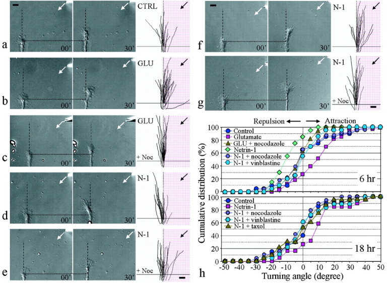
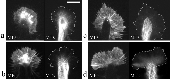
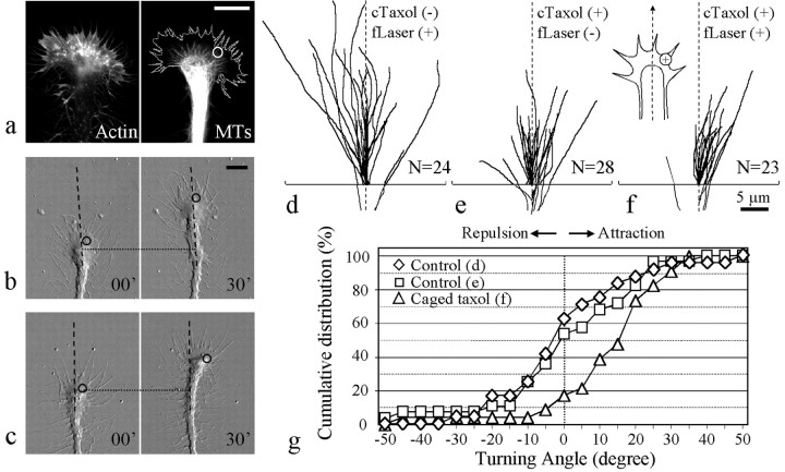
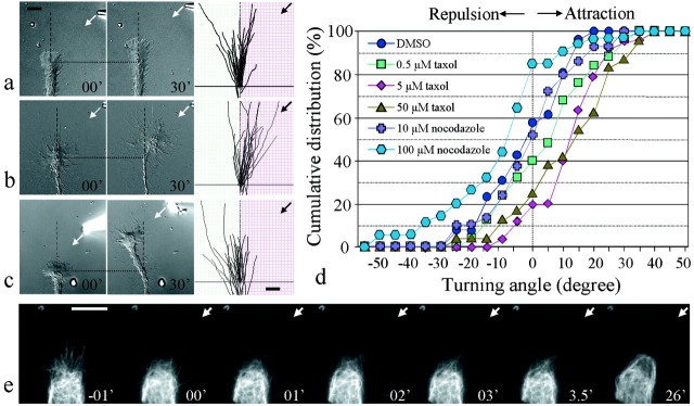
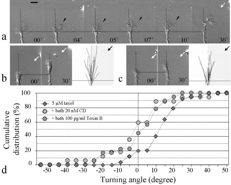
Pathfinding by nerve growth cones depends on attractive and repulsive turning in response to a variety of guidance cues. Here we present direct evidence to demonstrate an essential and instructive role for microtubules (MTs) in growth cone steering. First, both growth cone attraction and repulsion induced by diffusible cues in culture can be completely blocked by low concentrations of drugs that specifically inhibit dynamic microtubule ends in the growth cone. Second, direct focal photoactivated release of the microtubule-stabilizing drug taxol on one side of the growth cone consistently induces attraction (turning toward the site of application). Using the focal pipette application method, we also show that local MT stabilization by taxol induces growth cone attraction, whereas local MT destabilization by the microtubule-disrupting drug nocodazole induces repulsion (turning away). Finally, the microtubule-initiated attractive turning requires the participation of the actin cytoskeleton: local microtubule stabilization induces preferential protrusion of lamellipodia before the attractive turning, and the attraction can be abolished by inhibition of either actin polymerization or the Rho family GTPases. Together, these results demonstrate a novel steering mechanism for growth cones in which local and selective modification of dynamic microtubules can initiate and instruct directional steering. With the subsequent concerted activity of the actin cytoskeleton, this microtubule-initiated mechanism provides the growth cone with the additional means to efficiently navigate through its environment.


Figures

Fig. 1.
Fig. 1.
Blocking of growth cone turning responses to guidance gradients by inhibition of MT dynamics. a–e, Growth cone turning induced in 6 hr cultures. A gradient of glutamate (b) or netrin-1 (d) was applied to the growth cone by repetitive pulsatile pressure ejection of either 50 μm glutamate (GLU) or 5 μg/ml netrin-1 (N-1) solution from a micropipette (see Materials and Methods). The control (a) was done with the same pulsatile pipette application of culture medium (CTRL) to the growth cone. Images of the growth cone were collected at the onset (left image) and at the end (right image) of a 30 min application period. The origin is the center of the growth cone at the onset of the experiment.Dotted lines indicate corresponding positions along the neurite; dashed lines indicate the original direction of extension. Superimposed traces on theright (same magnifications as the growth cone images) depict the trajectory of neurite extension during the 30 min period for all of the growth cones in each group. Arrows indicate the direction of the gradient. Bath application of 10 nmnocodazole (+ Noc) abolished both attraction induced by glutamate (c) and repulsion induced by netrin-1 (e). f, g, Netrin-1-induced growth cone attraction in 18 hr cultures (f) was also blocked by the presence of 10 nm nocodazole in the bath (g). Scale bars: a–g, 10 μm. h, The distribution of turning angles is presented to depict the overall responses under different experimental conditions. For each condition, the percentage value refers to the percentage of growth cones with the turning angle less than or equal to a given angular value. Data shown are turning responses induced by glutamate and netrin-1 in 6 hr (top panel) and 18 hr (bottom panel) cultures without and with the presence of the MT drugs nocodazole and vinblastine in bath.
Fig. 2.
Fig. 2.
Double fluorescent imaging of microtubules and actin cytoskeleton in growth cones exposed to different MT-specific drugs. a, Representative pair of fluorescent images of the actin microfilaments (MFs) and MTs of a control growth cone treated with medium only. b–d, Representative pairs of MT and MF images of Xenopusgrowth cones treated with (in nm) 7 taxol (b), 10 nocodazole (c), and 4 vinblastine (d). All of the cells were fixed at 20 min after the treatment. For clarity, the margin of the growth cone lamellipodia in MT staining has been outlined. Scale bar, 10 μm.
Fig. 3.
Fig. 3.
Attractive turning of Xenopusgrowth cones induced by repetitive FLIP of caged taxol.a, Immunostaining of the actin cytoskeleton and microtubules in the growth cone. For clarity, the margin of the growth cone lamellipodia in MT staining has been outlined. Thecircle indicates the location of the laser irradiation.b, Control growth cone (no caged taxol) at the beginning and end of 30 min of repetitive laser irradiation;circles indicate the laser spot. The dotted line indicates corresponding positions along the neurite;dashed lines indicate the original direction of extension. c, Growth cone loaded with caged taxol at the onset and end of 30 min of repetitive FLIP. Numbers inb and c represent minutes after the onset of repetitive laser irradiation. d–f, Superimposedtraces of growth cone extension during the 30 min experimental period in two control groups [d, focal laser (fLaser) on but without caged taxol (cTaxol); e, laser off but with caged taxol] and in the group exposed to repetitive FLIP of caged taxol (f). g, The cumulative distribution illustrates the overall turning responses of all of the growth cones examined in these three groups. Scale bars:a–c, 10 μm; f, 5 μm.
Fig. 4.
Fig. 4.
Turning responses induced by focal application of MT-specific drugs through a micropipette. a, In the control, growth cones were exposed to focal application of 1% DMSO. Representative images of a control growth cone at the onset (left image) and at the end (right image) of the 30 min application are shown together with the superimposedtraces of all of the growth cones (same magnifications as the growth cone images). See the legend to Figure 1 for details.b, Attractive turning induced by focal application of 5 μm taxol. c, Repulsive turning induced by focal application of 100 μm nocodazole.Arrows in a–c indicate the direction of the gradient. d, Overall responses ofXenopus growth cones to focal application of taxol and nocodazole are shown in the cumulative distribution of turning angles.e, Live imaging sequence of rhodamine-labeled MTs in a growth cone exposed to focal taxol application. Arrowsindicate the direction of the focal taxol application.Numbers represent times after the onset of taxol application, with the first frame acquired 1 min before the onset of taxol application. Scale bars, 10 μm.
Fig. 5.
Fig. 5.
Involvement of the actin cytoskeleton in MT-initiated growth cone turning. a, Representative time lapse sequence of the attractive turning response of aXenopus growth cone induced by focal taxol application. Note the increased protrusion of lamellipodia on the side of the growth cone facing the pipette before the actual turning of the growth cone (arrowheads). b, Growth cone response to focal application of 5 μm taxol in the presence of 20 nm cytochalasin D in bath. Representative images of a growth cone at the onset (left image) and at the end (right image) of the 30 min taxol application are shown together with the superimposed traces of all of the growth cones on the right (same magnifications as the growth cone images). See the legend to Figure 1 for details.c, Growth cone response to focal application of 5 μm taxol in the presence of 100 pg/ml toxin B in bath. Scale bar, 10 μm. Arrows indicate the direction of the taxol gradient. d, Overall responses of growth cones to focal taxol application in the presence of toxin B (to inhibit the Rho GTPases) and cytochalasin D (to inhibit the actin assembly) are shown as the cumulative distribution of turning angles.

References

    1. Bentley D, O'Connor T. Cytoskeletal events in growth cone steering. Curr Opin Neurobiol. 1994;4:43–48. - PubMed
    1. Bentley D, Toroian-Raymond A. Disoriented pathfinding by pioneer neurone growth cones deprived of filopodia by cytochalasin treatment. Nature. 1986;323:712–715. - PubMed
    1. Bradke F, Dotti CG. The role of local actin instability in axon formation. Science. 1999;283:1931–1934. - PubMed
    1. Challacombe JF, Snow DM, Letourneau PC. Dynamic microtubule ends are required for growth cone turning to avoid an inhibitory guidance cue. J Neurosci. 1997;17:3085–3095. - PMC - PubMed
    1. Chang S, Rodionov VI, Borisy GG, Popov SV. Transport and turnover of microtubules in frog neurons depend on the pattern of axonal growth. J Neurosci. 1998;18:821–829. - PMC - PubMed

Publication types

MeSH terms