. 2002 Nov;14(11):2659-79.
doi: 10.1105/tpc.006155.

The Chlamydomonas reinhardtii plastid chromosome: islands of genes in a sea of repeats

Affiliations

The Chlamydomonas reinhardtii plastid chromosome: islands of genes in a sea of repeats

Jude E Maul et al. Plant Cell. 2002 Nov.

Abstract

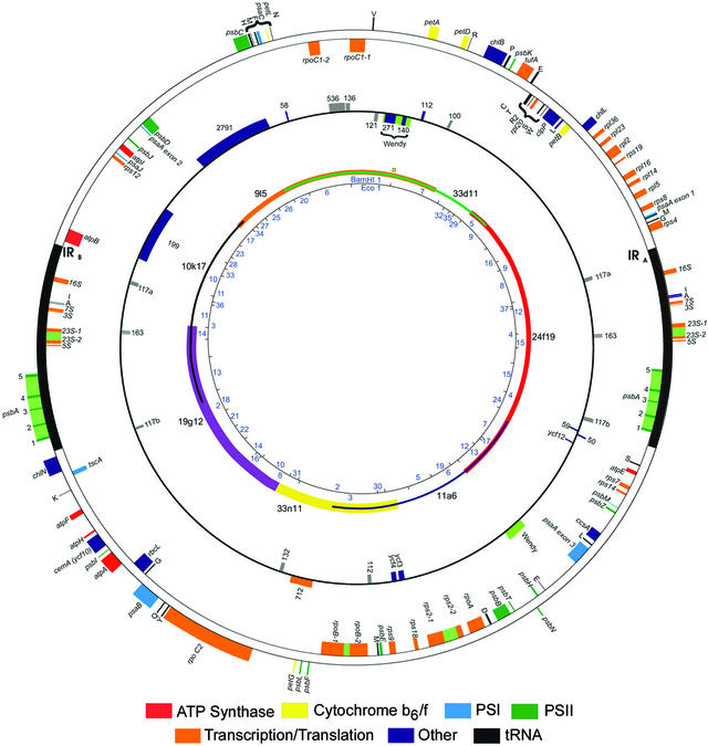
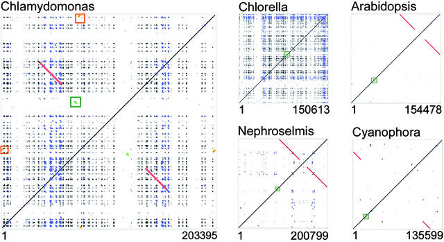
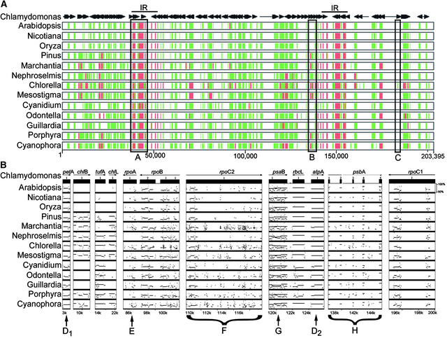
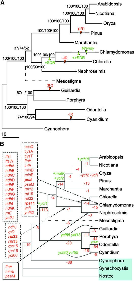
Chlamydomonas reinhardtii is a unicellular eukaryotic alga possessing a single chloroplast that is widely used as a model system for the study of photosynthetic processes. This report analyzes the surprising structural and evolutionary features of the completely sequenced 203,395-bp plastid chromosome. The genome is divided by 21.2-kb inverted repeats into two single-copy regions of approximately 80 kb and contains only 99 genes, including a full complement of tRNAs and atypical genes encoding the RNA polymerase. A remarkable feature is that >20% of the genome is repetitive DNA: the majority of intergenic regions consist of numerous classes of short dispersed repeats (SDRs), which may have structural or evolutionary significance. Among other sequenced chlorophyte plastid genomes, only that of the green alga Chlorella vulgaris appears to share this feature. The program MultiPipMaker was used to compare the genic complement of Chlamydomonas with those of other chloroplast genomes and to scan the genomes for sequence similarities and repetitive DNAs. Among the results was evidence that the SDRs were not derived from extant coding sequences, although some SDRs may have arisen from other genomic fragments. Phylogenetic reconstruction of changes in plastid genome content revealed that an accelerated rate of gene loss also characterized the Chlamydomonas/Chlorella lineage, a phenomenon that might be independent of the proliferation of SDRs. Together, our results reveal a dynamic and unusual plastid genome whose existence in a model organism will allow its features to be tested functionally.


Figures

Figure 1.
Figure 1.
The Plastid Chromosome of C. reinhardtii. The inner circle shows BamHI and EcoRI restriction fragments mapped according to Rochaix (1980) and numbered according to Grant et al. (1980). Position 0 is shown by an orange square near the 12 o'clock position. The second concentric circle indicates seven overlapping BAC clones that span the genome. The third circle shows genes and ORFs of unknown function, including those for which disruption experiments were unsuccessful. Gray boxes represent newly identified ORFs. The outer circle shows genes of known or presumed function, with sequenced or hypothesized (rps2; see text) introns shown in olive green. Genes are color coded by function, as shown at bottom.
Figure 2.
Figure 2.
Whole Chloroplast Genome Self-Comparison Dot Plots Generated by PipMaker. Dots off the main diagonal represent alignments with >50% identity. Structural features off the main diagonal are color coded as follows: red, inverted repeats; blue, small dispersed repeats in C. reinhardtii that also are detected in C. vulgaris and N. olivacea; green, psaA/psaB genes; orange, Wendy elements. Accession numbers for the cpDNA sequences are given at the end of Methods.
Figure 3.
Figure 3.
Cytogenomic Analyses of C. reinhardtii cpDNA. (A) Linear cpDNA fiber with two genome copies. The green signal represents the entire plastid genome, and the three red signals result from the hybridization of clones P-14 and P-16, which span a 10-kb portion of the inverted repeat. (B) Two monomeric cpDNA fibers visible in the same field of view. (C) Chloroplast DNA dimer of ∼180 μm. (D) A field showing various linear cpDNA fibers. Bars = 10 μm.
Figure 4.
Figure 4.
MultiPipMaker Analysis of 14 Sequenced Plastid Genomes, Illustrating Similarities and Differences between Chlamydomonas and Other Photosynthetic Plastid Genomes. (A) The reference genome, C. reinhardtii (top), was analyzed against 13 other species. Genes and their orientations are indicated by horizontal arrows, and the large inverted repeat (IR) is shown as a line. Positions on the linearized genome are shown across the bottom. The species for aligned genomes are shown at left, with accession numbers given at the end of Methods. Where alignments with the Chlamydomonas sequence were made, they are shown as 50 to 75% identity (green) or 75 to 100% identity (red). The boxes labeled A, B, and C denote features discussed in the text. (B) Selected regions were extracted from the analysis shown in (A), with aligned regions displayed graphically as horizontal lines drawn to show average percentage identity between 50 and 100% (at right of Arabidopsis plot). Positions of the selected sequences in the Chlamydomonas chloroplast genome are shown at bottom. The genes or braced areas labeled D to H denote features discussed in the text.
Figure 5.
Figure 5.
Plastid Genome Phylogeny with Changes in Gene Content and Structural Features for 14 Fully Sequenced Plastid Genomes and 2 Cyanobacterial Genomes. (A) Maximum likelihood phylogeny for 39 concatenated proteins totaling 8856 amino acids after removal of gaps and regions of ambiguous alignment. The tree is drawn with branch lengths reconstructed under maximum likelihood with the JTT model. Bootstrap support values are shown for nodes with 50% or greater support in maximum parsimony/neighbor joining/maximum likelihood RELL BP analyses. The dotted branch leading to Mesostigma indicates the uncertainty of the branching position for this taxon (see text). Arrows highlight noteworthy structural changes, including loss (−IR) or near loss (IR) of one copy of the inverted repeat, invasion by SDRs (+SDR) and their further proliferation (++SDR), and invasion by the Wendy element in Chlamydomonas. (B) Unambiguous gene losses (in red) and gains (in green) on each branch, assuming that a gene with a detected homolog in cyanobacterial outgroups (Synechocystis or Nostoc; shaded) was present in the ancestral plastid genome. Each gene gain is indicated; specific gene losses are shown for green algal branches only. Losses inferred to have occurred only once throughout the plastome phylogeny are shown in boldface; all other losses are inferred in multiple plastome lineages (see supplemental data online for mapping of all gene changes on every branch).
Figure 6.
Figure 6.
RNA Gel Blot Analysis for Selected PEP Genes and rps2. Total RNA (40 μg) from CC-125 cells grown in continuous light was analyzed by filter hybridization using 32P-labeled probes derived by PCR from rpoA and rpoC2 (A and C2, respectively). Probes corresponding to the rps2 ORFs were used in the lanes marked rps2-1 (previously ORF570) and rps2-2 (ORF208). Individual transcript sizes are noted and were estimated by comparison with RNA markers (Invitrogen) and the 16S rRNA hybridization control (16S). The symbols mark transcripts that are discussed in the text, generally those considered most likely to encode the protein product based on size or on hybridization with two probes from the same gene.
Figure 7.
Figure 7.
RT-PCR for Genes That Encode PEP Subunits. Approximately 1 μg of total RNA from light-grown CC-125 cells was subjected to a 1-h reverse transcription reaction, followed by 30 cycles of PCR using gene-specific primers (Lilly et al., 2002). At left, RT-PCR products for an internal region of each PEP gene. Lanes are as follows: M, molecular size markers; A, rpoA; C2, rpoC2; C1a, rpoC1a; C1b, rpoC1b; B1, rpoB1; and B2, rpoB2. At right, RT-PCR products spanning the rpoC2 gene. Lanes correspond to regions of the 3119–amino acid rpoC2 ORF, as indicated by the diagram below the gel. Open bars represent regions that were amplified by RT-PCR, and closed bars represent regions not amplified by RT-PCR.
Figure 8.
Figure 8.
BOXSHADE Alignments of Newly Identified PEP Coding Regions. (A) Multiple sequence alignment of the translated rpoA sequence of C. reinhardtii with the five most similar peptide sequences, as determined by tBLASTX analysis. (B) A section of the multiple sequence alignment of the translated rpoC1 sequence from C. reinhardtii with those of other related species. This excerpt highlights the zinc binding domain (conserved Cys residues are denoted by asterisks), which is found in most prokaryote-like rpoC1 proteins and implicated in transcription termination. Full names and accession numbers are given at the end of Methods.

References

    1. Adachi, J., and Hasegawa, M. (1996). MOLPHY version 2.3: Programs for molecular phylogeny based on maximum likelihood. In Computer Science Monographs 28. (Tokyo: Institute of Statistical Mathematics).
    1. Allison, L.A. (2000). The role of sigma factors in plastid transcription. Biochimie 82, 537–548. - PubMed
    1. Altschul, S.F., Madden, T.L., Schaffer, A.A., Zhang, J., Zhang, Z., Miller, W., and Lipman, D.J. (1997). Gapped BLAST and PSI-BLAST: A new generation of protein database search programs. Nucleic Acids Res. 25, 3389–3402. - PMC - PubMed
    1. Backert, S., Dorfel, P., and Börner, T. (1995). Investigation of plant organellar DNAs by pulsed-field gel electrophoresis. Curr. Genet. 28, 390–399. - PubMed
    1. Barkan, A., and Goldschmidt-Clermont, M. (2000). Participation of nuclear genes in chloroplast gene expression. Biochimie 82, 559–572. - PubMed

Publication types

MeSH terms

Associated data

LinkOut - more resources