. 2002 Nov;14(11):2707-22.
doi: 10.1105/tpc.005884.

VH1, a provascular cell-specific receptor kinase that influences leaf cell patterns in Arabidopsis

Affiliations

VH1, a provascular cell-specific receptor kinase that influences leaf cell patterns in Arabidopsis

Nicole K Clay et al. Plant Cell. 2002 Nov.

Abstract

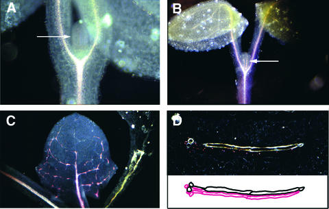
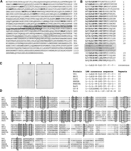
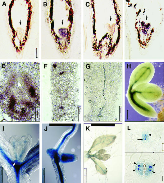
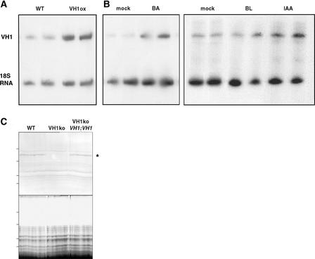
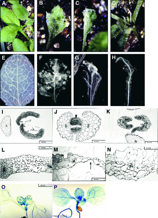
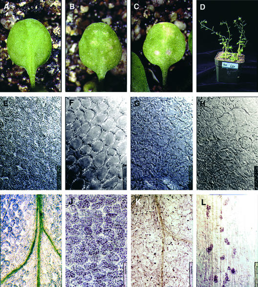
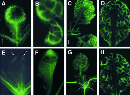
The formation of the venation pattern in leaves is ideal for examining signaling pathways that recognize and respond to spatial and temporal information, because the pattern is two-dimensional and heritable and the resulting veins influence the three-dimensional spatial organization of the surrounding differentiating leaf cell types. We identified a provascular/procambial cell-specific gene that encodes a Leu-rich repeat receptor kinase, which we named VASCULAR HIGHWAY1 (VH1). A change in the expression domain and level of VH1 marks the transition from an uncommitted provascular state to a committed procambial state in early vascular development. The coding sequence, expression pattern, and transgenic phenotypes together suggest that VH1 transduces extracellular spatial and temporal signals into downstream cell differentiation responses in provascular/procambial cells.


Figures

Figure 1.
Figure 1.
VH1 Is Associated with a PV/PC Enhancer. (A) and (B) Seedlings from an enhancer trap line at successive stages of leaf development were stained for GUS expression and viewed under dark-field illumination (GUS staining appears pink). Arrows point to GUS-stained leaf primordium (A) and to a GUS-stained PC strand of the midvein (B). (C) GUS staining is specific to PC cells, and when those cells mature, they no longer stain for GUS activity. (D) A transverse section through an expanding leaf shows a single file of GUS-stained PC cells underneath the xylem strand (top). Below is the corresponding tracing of xylem cells (black) and PC cells (pink).
Figure 2.
Figure 2.
VH1 Encodes a LRR Receptor Kinase. (A) The predicted VH1 protein contains the following regions: a signal peptide (amino acids 1 to 31); a putative Leu zipper (italicized); an extracellular LRR domain (amino acids 105 to 708) that contains a buried 68–amino acid island (underlined and in boldface) and is flanked by two conservatively spaced Cys-pair regions (underlined); a transmembrane domain (italicized and underlined) that is flanked by stop transfer sequences; and an intracellular kinase domain (amino acids 795 to 1143) with Ser/Thr specificity. Potential N-glycosylation sites are shown in boldface. (B) The extracellular domain contains 22 LRRs with a unit length of 24 amino acids. Numbers at right of the LRR domain indicate the specific LRR number, and the bottom line is the consensus sequence for the VH1 LRR. Dashes stand for any amino acid, and φ indicates an aliphatic amino acid residue. Amino acid residues that match the deduced consensus sequence are shown in boldface. Lowercase letters in the consensus sequence indicate residues identical in at least half of the repeats. A 68–amino acid island that does not fit the consensus sequence is buried between the 18th and 19th LRRs (black box). In the table at bottom, the LRR consensus sequence of VH1 is compared with those of other LRR-containing signaling proteins: Arabidopsis BRI1, CLV1, HAESA, and ERECTA; rice Xa21; tomato Cf-9; and Drosophila Toll. (C) Maltose binding protein (MBP) fusions to an active and an inactive version of the VH1 kinase domain (MBP::VH1 and MBP::K866N, respectively) were affinity purified from isopropylthio-β-galactoside–induced E. coli cells and used in an in vitro kinase assay. The gel at right is a Coomassie blue–stained 10% SDS-PAGE gel, and the gel at left is the corresponding autoradiogram, which shows 32P-labeled MBP::VH1 (lane 2) as a product of an autophosphorylation reaction. MBP::K866N is in lane 1. Lines at left indicate molecular mass markers (107, 76, and 52 kD from top to bottom). (D) Alignment of the kinase domain among putative LRR receptor kinases in plants. Residues that are conserved among all of the compared sequences are boxed, and those that are conserved among at least four sequences are shaded. The 12 conserved protein kinase domains are indicated I to XI (Hanks and Quinn, 1991). The 15 invariant amino acids present in all protein kinases are indicated by asterisks.
Figure 3.
Figure 3.
VH1 Expression Is Associated with PV/PC Sites. (A) to (G) Digoxigenin-labeled VH1-specific RNA probes were hybridized to serial sections of tissue from wild-type plants. Transverse sections of young leaves ([F] and [G]) as well as longitudinal sections of globe-stage ([A] and [B]) and heart-stage ([C] and [D]) embryos (arrows) and leaf primordia (E) were hybridized to antisense ([B] and [D] to [F]) and sense ([A], [C], and [G]) probes. (A) to (D) are at the same magnification; bar in (A) = 50 μm. Bars in (F) to (G) = 25 μm. (H) to (L) Transgenic plants containing the VH1::GUS reporter construct were stained for GUS activity, and whole-mounts ([H] to [K]) and serial transverse sections through a young root (K) were viewed using Nomarski optics. Whole-mount tissues include bent-cotyledon-stage embryos (H), leaf primordia (arrow) and very young leaves (I), lateral root primordia (J), and young cauline leaves that subtend floral buds (K). Bars = 50 μm for (H) and 40 μm for (I) to (K). In (L), arrows indicate PC cells that will differentiate as xylem, and arrowheads indicate PC cells that will differentiate as phloem. Bars = 8 μm.
Figure 4.
Figure 4.
VH1 Expression Data from Transgenic Lines and from Exogenous Application of Hormones. (A) and (B) Both VH1 (425-bp fragment) and 18S RNA (315 bp) were amplified by PCR from cDNA generated from total RNA for each sample and loaded on a denaturing acrylamide gel in duplicate. The VH1 band was quantified relative to the 18S RNA. (A) One-week-old wild-type (WT) and 35S CaMV::VH1 (VH1ox) transgenic seedlings. (B) Etiolated seedlings were incubated for 1 h in a salt solution alone (mock) or with the addition of benzyladenine (BA), 24-epibrassinolide (BL), or indoleacetic acid (IAA). (C) The gel at top shows a protein blot of whole-cell extracts from 5-day-old seedlings probed with VH1 antiserum. Both the wild type (WT) and the vh1 insertion line containing a wild-type VH1 transgene driven by its own promoter (VH1ko + VH1::VH1) produced an ∼150-kD band, whereas the vh1 insertion line alone (VH1ko) did not. The gel at bottom shows the corresponding Coomassie blue–stained 7.5% SDS-PAGE gel demonstrating that the samples were loaded equally. The asterisk indicates the VH1 protein band. Lines at left indicate molecular mass markers (184, 121, 86, and 69 kD from top to bottom).
Figure 5.
Figure 5.
Ectopic VH1 Expression Results in Premature Leaf Cell Differentiation. Juvenile leaves from wild-type plants were used for comparison ([A], [E], [I], and [L]). (A) to (H) Fully expanded juvenile leaves were viewed with a dissecting microscope ([A] to [D]) and then were cleared and viewed using dark-field optics ([E] to [H]) to visualize the venation pattern and the mesophyll cell layers. (I) to (N) Transverse sections through leaf primordia ([I] and [J]) and expanding (K) and fully expanded ([L] to [N]) juvenile leaves were stained with toluidine blue and viewed using bright-field optics. The arrowhead in (J) points to primordial ground cells (normally identified by their dense cytoplasmic staining) that have prematurely ceased cell division and have differentiated, resulting in leaves with fewer mesophyll cells that are unusually sparse in interveinal regions (K). (L) to (N) are oriented so that the adaxial surface of the leaf section is at top. The adaxial surface is above the xylem (light blue) portion of the midvein. The arrow in (M) points to a region composed of a single epidermal layer. Bars = 50 μm for (I) and (K) to (M) and 40 μm for (J) and (N). (O) and (P) Plants containing both the 35S CaMV::VH1 and the VH1::GUS constructs (P) expressed GUS constitutively, expanding the GUS expression domain seen in plants containing only the VH1::GUS reporter construct (O). Bars = 400 μm.
Figure 6.
Figure 6.
Loss of VH1 Results in Premature Leaf Senescence and Possible Defect in Vascular Transport. Juvenile leaves from wild-type (A) and vh1 mutant ([B] and [C]) 3-week-old plants were viewed with a dissecting microscope to visualize senescence. In (D), vh1 mutant plants are at left and wild-type plants are at right. Expanding ([E] and [G]) and fully expanded ([F] and [H]) leaves of wild-type ([E] and [F]) and vh1 mutant ([G] and [H]) plants were cleared and viewed with Nomarski optics to visualize the chloroplast-bearing mesophyll cells. Bars = 25 μm for (A) to (H). Cleared leaves of wild-type ([I] and [J]) and vh1 mutant ([K] and [L]) plants were stained with I/KI for starch accumulation. Bars = 50 μm for (I) and (K) and 25 μm for (J) and (L).
Figure 7.
Figure 7.
Loss of VH1 Results in Defective Vascular Transport of a Phloem-Mobile Dye Early in Leaf Development. The pattern of phloem transport of CF was imaged in the sink leaves of wild-type ([A] to [D]) and vh1 mutant ([E] to [H]) plants at successive stages of leaf development. Arrows in (E) indicate top portions of major veins that are fluorescing. (A), (B), (E), and (F) are at the same magnification; bar in (A) = 40 μm. (C), (D), (G), and (H) are at the same magnification; bar in (C) = 400 μm.

References

    1. Badenhuizen, N.P. (1969). The Biogenesis of Starch Granules in Higher Plants. (New York: Appleton-Century-Crofts).
    1. Baima, S., Nobili, F., Sessa, G., Luchetti, S., Ruberti, I., and Morelli, G. (1995). The expression of the Athb-8 homeobox gene is restricted to provascular cells in Arabidopsis thaliana. Development 121, 4171–4182. - PubMed
    1. Baker, B., Zambryski, P., Staskawicz, B., and Dinesh-Kumar, S.P. (1997). Signaling in plant-microbe interactions. Science 276, 726–733. - PubMed
    1. Berleth, T., and Jurgens, G. (1993). The role of the monopteros gene in organizing the basal body region of the Arabidopsis embryo. Development 118, 575–587.
    1. Berleth, T., Mattsson, J., and Hardtke, C.S. (2000). Vascular continuity and auxin signals. Trends Plant Sci. 5, 387–393. - PubMed

Publication types

MeSH terms