

Positioning of nuclei in Arabidopsis root hairs: an actin-regulated process of tip growth
- PMID: 12417712
- PMCID: PMC152738
- DOI: 10.1105/tpc.005892
Positioning of nuclei in Arabidopsis root hairs: an actin-regulated process of tip growth
Abstract
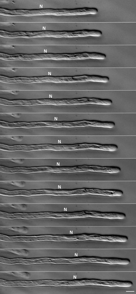
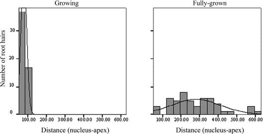
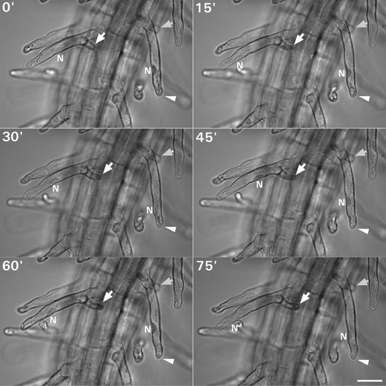
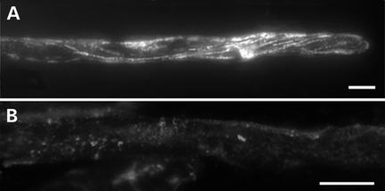
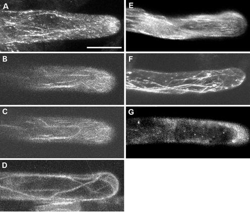
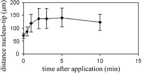
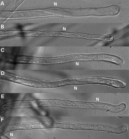
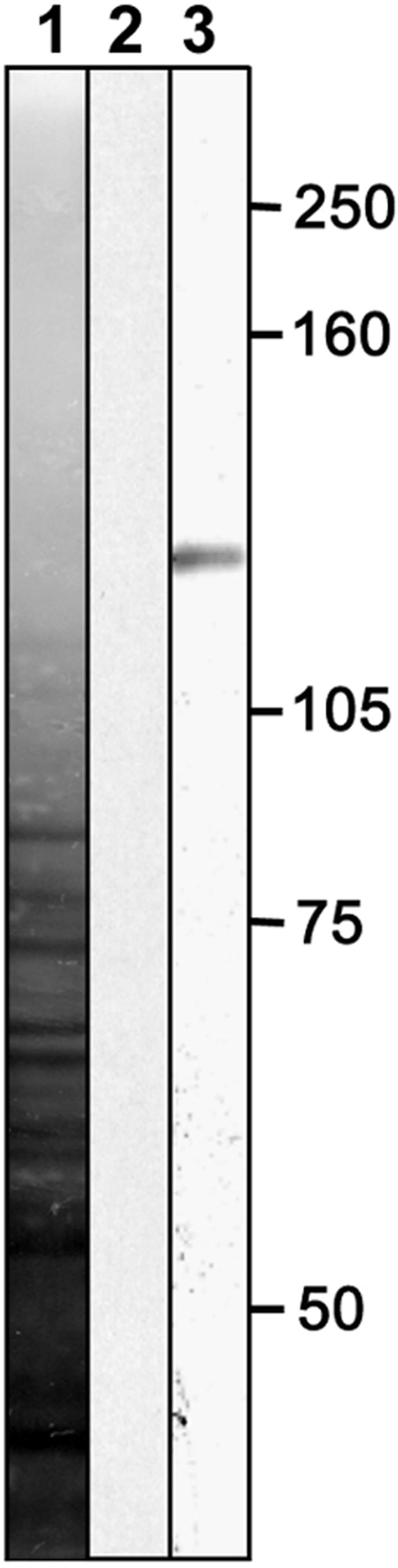
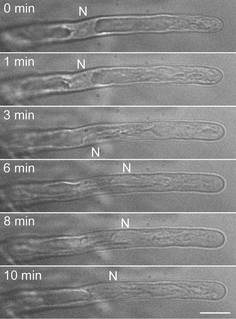
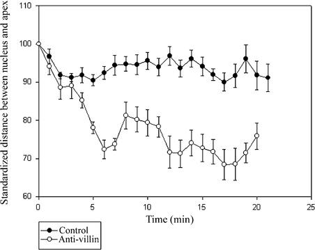
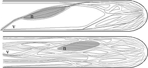
In growing Arabidopsis root hairs, the nucleus locates at a fixed distance from the apex, migrates to a random position during growth arrest, and moves from branch to branch in a mutant with branched hairs. Consistently, an artificial increase of the distance between the nucleus and the apex, achieved by entrapment of the nucleus in a laser beam, stops cell growth. Drug studies show that microtubules are not involved in the positioning of the nucleus but that subapical fine F-actin between the nucleus and the hair apex is required to maintain the nuclear position with respect to the growing apex. Injection of an antibody against plant villin, an actin filament-bundling protein, leads to actin filament unbundling and movement of the nucleus closer to the apex. Thus, the bundled actin at the tip side of the nucleus prevents the nucleus from approaching the apex. In addition, we show that the basipetal movement of the nucleus at root hair growth arrest requires protein synthesis and a functional actin cytoskeleton in the root hair tube.
Figures
Similar articles
-
PCaP2 regulates nuclear positioning in growing Arabidopsis thaliana root hairs by modulating filamentous actin organization.Plant Cell Rep. 2015 Aug;34(8):1317-30. doi: 10.1007/s00299-015-1789-6. Epub 2015 May 1. Plant Cell Rep. 2015. PMID: 25929794
-
Nuclear dynamics and programmed cell death in Arabidopsis root hairs.Plant Sci. 2016 Dec;253:77-85. doi: 10.1016/j.plantsci.2016.08.006. Epub 2016 Oct 1. Plant Sci. 2016. PMID: 27968999
-
ACTIN2 is essential for bulge site selection and tip growth during root hair development of Arabidopsis.Plant Physiol. 2002 Aug;129(4):1464-72. doi: 10.1104/pp.005777. Plant Physiol. 2002. PMID: 12177460 Free PMC article.
-
Building a hair: tip growth in Arabidopsis thaliana root hairs.Philos Trans R Soc Lond B Biol Sci. 2002 Jun 29;357(1422):815-21. doi: 10.1098/rstb.2002.1092. Philos Trans R Soc Lond B Biol Sci. 2002. PMID: 12079677 Free PMC article. Review.
-
The actin cytoskeleton in root hairs: all is fine at the tip.Curr Opin Plant Biol. 2013 Dec;16(6):749-56. doi: 10.1016/j.pbi.2013.10.003. Curr Opin Plant Biol. 2013. PMID: 24446547 Review.
Cited by
-
Root hair growth: it's a one way street.F1000Prime Rep. 2015 Feb 3;7:23. doi: 10.12703/P7-23. eCollection 2015. F1000Prime Rep. 2015. PMID: 25750741 Free PMC article. Review.
-
Micromanipulation of amyloplasts with optical tweezers in Arabidopsis stems.Plant Biotechnol (Tokyo). 2020 Dec 25;37(4):405-415. doi: 10.5511/plantbiotechnology.20.1201a. Plant Biotechnol (Tokyo). 2020. PMID: 33850427 Free PMC article.
-
Arabidopsis VILLIN1 generates actin filament cables that are resistant to depolymerization.Plant Cell. 2005 Feb;17(2):486-501. doi: 10.1105/tpc.104.028555. Epub 2005 Jan 19. Plant Cell. 2005. PMID: 15659626 Free PMC article.
-
Cytoskeleton dynamics control the first asymmetric cell division in Arabidopsis zygote.Proc Natl Acad Sci U S A. 2016 Dec 6;113(49):14157-14162. doi: 10.1073/pnas.1613979113. Epub 2016 Nov 22. Proc Natl Acad Sci U S A. 2016. PMID: 27911812 Free PMC article.
-
Polarization of the endomembrane system is an early event in fucoid zygote development.BMC Plant Biol. 2006 Feb 23;6:5. doi: 10.1186/1471-2229-6-5. BMC Plant Biol. 2006. PMID: 16504093 Free PMC article.
References
-
- Andersland, J.M., Fisher, D.D., Wymer, C.L., Cyr, R.J., and Parthasarathy, M.V. (1994). Characterization of a monoclonal antibody prepared against plant actin. Cell Motil. Cytoskeleton 29, 339–344. - PubMed
-
- Anthony, R.G., Waldin, T.R., Ray, J.A., Bright, S.W., and Hussey, P.J. (1998). Herbicide resistance caused by spontaneous mutation of the cytoskeletal protein tubulin. Nature 393, 260–263. - PubMed
-
- Baluška, F., Salaj, J., Mathur, J., Braun, M., Jasper, F., Šamaj, J., Chua, N.-H., Barlow, P.W., and Volkman, D. (2000). Root hair formation: F-actin-dependent tip growth is initiated by local assembly of profilin-supported F-actin meshworks accumulated within expansin-enriched bulges. Dev. Biol. 227, 618–632. - PubMed
-
- Bibikova, T.N., Blancaflor, E.B., and Gilroy, S. (1999). Microtubules regulate tip growth and orientation in root hairs of Arabidopsis thaliana. Plant J. 17, 657–665. - PubMed
MeSH terms
Substances
LinkOut - more resources
Full Text Sources
Other Literature Sources
Miscellaneous
