. 2002 Dec;22(23):8114-21.
doi: 10.1128/MCB.22.23.8114-8121.2002.

Identification of a human decapping complex associated with hUpf proteins in nonsense-mediated decay

Affiliations

Identification of a human decapping complex associated with hUpf proteins in nonsense-mediated decay

Jens Lykke-Andersen. Mol Cell Biol. 2002 Dec.

Abstract

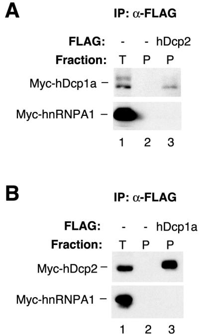
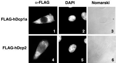
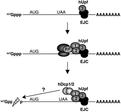
Decapping is a key step in general and regulated mRNA decay. In Saccharomyces cerevisiae it constitutes a rate-limiting step in the nonsense-mediated decay pathway that rids cells of mRNAs containing premature termination codons. Here two human decapping enzymes are identified, hDcp1a and hDcp2, as well as a homolog of hDcp1a, termed hDcp1b. Transiently expressed hDcp1a and hDcp2 proteins localize primarily to the cytoplasm and form a complex in human cell extracts. hDcp1a and hDcp2 copurify with decapping activity, an activity sensitive to mutation of critical hDcp residues. Importantly, coimmunoprecipitation assays demonstrate that hDcp1a and hDcp2 interact with the nonsense-mediated decay factor hUpf1, both in the presence and in the absence of the other hUpf proteins, hUpf2, hUpf3a, and hUpf3b. These data suggest that a human decapping complex may be recruited to mRNAs containing premature termination codons by the hUpf proteins.


Figures

FIG. 1.
FIG. 1.
Human homologs of S. cerevisiae decapping enzymes. (A) Schematics of S. cerevisiae Dcp1p (yDcp1) and the human homologs hDcp1a and hDcp1b. (B) Schematics of S. cerevisiae Dcp2p (yDcp2) and the human homolog hDcp2. Gray bars indicate regions of similarity (percent identity given below) between human and S. cerevisiae Dcps. The black bars in yDcp2 and hDcp2 indicate the conserved mutT domains. Residues critical for decapping activity are indicated (see Fig. 4). aa, amino acids.
FIG. 2.
FIG. 2.
hDcp1a and hDcp2 interact in cell extracts. (A) Western blot for Myc-tagged hDcp1a and hnRNPA1 proteins, which were transiently expressed in HEK293 cells together with an empty vector (lanes 1 and 2) or a plasmid encoding FLAG-tagged hDcp2 (lane 3) and subjected to anti-FLAG immunoprecipitation after RNase treatment. (B) Western blot for Myc-tagged hDcp2 and hnRNPA1 proteins, transiently expressed in HEK293 cells together with an empty vector (lanes 1 and 2) or a plasmid encoding FLAG-tagged hDcp1a (lane 3) and subjected to anti-FLAG immunoprecipitation after RNase treatment. Immunoprecipitates (P; lanes 2 and 3) are compared to 5% of total extract (T; lane 1).
FIG. 3.
FIG. 3.
Decapping assays with immunopurified and recombinant hDcp1a and hDcp2. α-32P-m7cap-labeled RNA was incubated with immunopurified FLAG-tagged hDcp1a (lanes 4, 5, 9, and 10; corresponding to 0.2 × 10⁶, 1 × 10⁶, 1 × 10⁶, and 1 × 10⁶ cells' worth of protein, respectively), hDcp2 (lanes 6 and 7; 1 × 10⁶ and 5 × 10⁶ cells' worth, respectively), mock-purified cell extract (lanes 2 and 3; 1 × 10⁶ and 5 × 10⁶ cells' worth, respectively), or no cell extract (lanes 1, 8, and 11) or with purified bacterially expressed GST (lanes 12 and 13; 10 and 100 ng, respectively), GST-hDcp1a (lanes 14 and 15; 10 and 100 ng, respectively), GST-hDcp2 (lanes 16 and 17; 10 and 100 ng, respectively), or GST-hDcp1a and GST-hDcp2 (lanes 18 and 19; 10 and 100 ng each, respectively), and products were separated by thin-layer chromatography. The reaction mixture in lane 10 was treated with nucleoside diphosphate kinase (NDPK) and ATP. The migration of unlabeled GMP, GDP, m7GMP, m7GDP, and m7GTP (25 μg) is indicated on the left.
FIG. 4.
FIG. 4.
Mutation of critical residues in hDcp1a and hDcp2 disrupts decapping activity. The figure shows the results of decapping assays with immunopurified wild-type and mutant hDcp1a and hDcp2 proteins as indicated. The amount of protein corresponds to 5 × 10⁶, 0.5 × 10⁶, 5 × 10⁶, 5 × 10⁶, 1 × 10⁶, 0.5 × 10⁶, 0.5 × 10⁶, 5 × 10⁶, and 5 × 10⁶ cells in lanes 2 to 10, respectively. The relative amount of m7GDP generated by each hDcp mutant was quantified with a phosphorimager and is given below relative to that of wild-type hDcps, which was set to 100%. A fraction of each assay (lanes 3 to 10) was subjected to Western blotting with anti-FLAG antibodies, shown in the lower panel. wt, wild type.
FIG. 5.
FIG. 5.
Transiently expressed hDcp1a and hDcp2 proteins localize primarily in the cytoplasm. The figure shows the results of immunocytochemical staining of fixed, permeabilized HeLa cells, transiently expressing FLAG-tagged hDcp1a (panels 1 to 3) or hDcp2 (panels 4 to 6). Cells were stained with anti-FLAG M2 monoclonal antibody and visualized with Texas red-conjugated anti-mouse immunoglobulin G antibody (panels 1 and 4). Nuclei are visualized with 4′,6′-diamidino-2-phenylindole (DAPI) (panels 2 and 5). Panels 1 to 6 show one cell, representative of at least 100 observed transfected cells.
FIG. 6.
FIG. 6.
hDcp1a and hDcp2 interact with the NMD protein hUpf1. (A) Western blot for Myc-tagged hDcp1a, hDcp2, or hnRNPA1, transiently expressed in HEK293 cells together with an empty vector (lanes 1 and 2) or a plasmid encoding FLAG-tagged hUpf1 (lane 3) and subjected to anti-FLAG immunoprecipitation after RNase treatment. (B) Western blot for Myc-tagged hUpf1 or hnRNPA1, transiently expressed in HEK293 cells together with an empty vector (lanes 1, 2, 4, and 5) or a plasmid encoding FLAG-tagged hDcp1a (lane 3) or hDcp2 (lane 6) and subjected to anti-FLAG immunoprecipitation after RNase treatment. Immunoprecipitates (P; lanes 2, 3, 5, and 6) are compared to 5% of the total extract (T; lanes 1 and 4) in panels A and B. (C) RNase-treated extracts from HEK293 cells transiently expressing Myc-hDcp1a, Myc-hnRNPA1, and FLAG-hUpf1 were immunodepleted with preimmune serum (pi; lanes 1, 2, and 3) or anti-hUpf1 (α1; lane 4), anti-hUpf2 (α2; lane 5), or anti-hUpf3b (α3b; lane 6) serum and subsequently immunoprecipitated with anti-FLAG antibody. Immunoprecipitates (upper two panels) were probed for the presence of Myc-hDcp1a and Myc-hnRNPA1 by Western blotting. Five percent of the total extract (T; lane 1) was compared to the pellets (P; lanes 2 to 5). Depleted extracts (D; lower five panels, lanes 3 to 6) were probed with monoclonal anti-Myc antibody or polyclonal rabbit antibodies against hUpf1, hUpf2, hUpf3a, and hUpf3b as indicated.
FIG. 7.
FIG. 7.
Model for how the interaction between hUpf1 and hDcp proteins may play a role in NMD. The EJC is deposited onto the mRNA upstream of exon-exon junctions after pre-mRNA splicing. hUpf3 associates with the EJC, followed by hUpf2 and hUpf1. A translation termination event upstream of the EJC/hUpf complex may allow hUpf1 to recruit the hDcp proteins to trigger mRNA decapping. RF, translation release factor.

References

    1. Allmang, C., E. Petfalski, A. Podtelejnikov, M. Mann, D. Tollervey, and P. Mitchell. 1999. The yeast exosome and human PM-Scl are related complexes of 3′→5′ exonucleases. Genes Dev. 13:2148-2158. - PMC - PubMed
    1. Bai, R. Y., C. Koester, T. Ouyang, S. A. Hahn, M. Hammerschmidt, C. Peschel, and J. Duyster. 2002. SMIF, a Smad4-interacting protein that functions as a co-activator in TGFβ signalling. Nat. Cell Biol. 4:181-190. - PubMed
    1. Barnes, C. A. 1998. Upf1 and Upf2 proteins mediate normal yeast mRNA degradation when translation initiation is limited. Nucleic Acids Res. 26:2433-2441. - PMC - PubMed
    1. Beelman, C. A., and R. Parker. 1995. Degradation of mRNA in eukaryotes. Cell 81:179-183. - PubMed
    1. Beelman, C. A., A. Stevens, G. Caponigro, T. E. LaGrandeur, L. Hatfield, D. M. Fortner, and R. Parker. 1996. An essential component of the decapping enzyme required for normal rates of mRNA turnover. Nature 382:642-646. - PubMed

MeSH terms