. 2002 Dec;22(23):8292-301.
doi: 10.1128/MCB.22.23.8292-8301.2002.

Esc1, a nuclear periphery protein required for Sir4-based plasmid anchoring and partitioning

Affiliations

Esc1, a nuclear periphery protein required for Sir4-based plasmid anchoring and partitioning

Erik D Andrulis et al. Mol Cell Biol. 2002 Dec.

Abstract

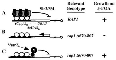
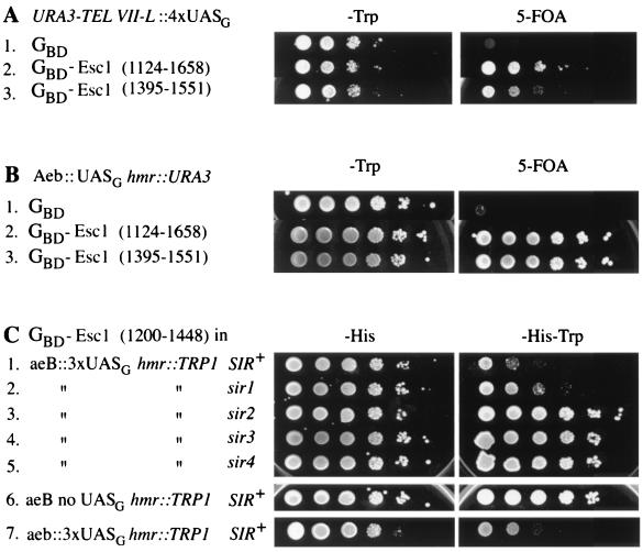
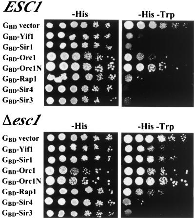
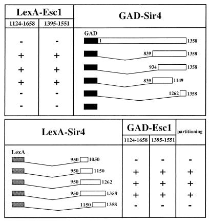
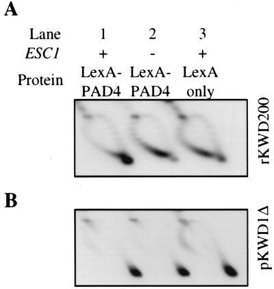
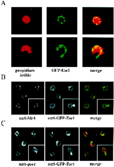
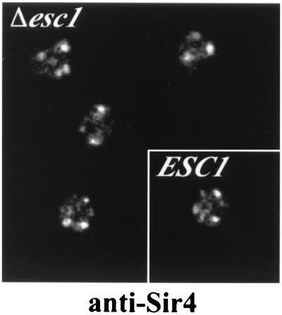
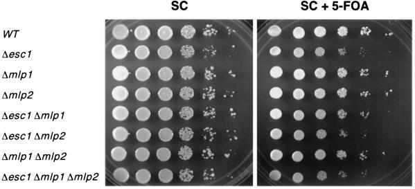
A targeted silencing screen was performed to identify yeast proteins that, when tethered to a telomere, suppress a telomeric silencing defect caused by truncation of Rap1. A previously uncharacterized protein, Esc1 (establishes silent chromatin), was recovered, in addition to well-characterized proteins Rap1, Sir1, and Rad7. Telomeric silencing was slightly decreased in Deltaesc1 mutants, but silencing of the HM loci was unaffected. On the other hand, targeted silencing by various tethered proteins was greatly weakened in Deltaesc1 mutants. Two-hybrid analysis revealed that Esc1 and Sir4 interact via a 34-amino-acid portion of Esc1 (residues 1440 to 1473) and a carboxyl-terminal domain of Sir4 known as PAD4 (residues 950 to 1262). When tethered to DNA, this Sir4 domain confers efficient partitioning to otherwise unstable plasmids and blocks the ability of bound DNA segments to rotate freely in vivo. Here, both phenomena were shown to require ESC1. Sir protein-mediated partitioning of a telomere-based plasmid also required ESC1. Fluorescence microscopy of cells expressing green fluorescent protein (GFP)-Esc1 showed that the protein localized to the nuclear periphery, a region of the nucleus known to be functionally important for silencing. GFP-Esc1 localization, however, was not entirely coincident with telomeres, the nucleolus, or nuclear pore complexes. Our data suggest that Esc1 is a component of a redundant pathway that functions to localize silencing complexes to the nuclear periphery.


Figures

FIG. 1.
FIG. 1.
Telomeric targeted silencing screen. A telomere with a URA3 reporter gene and four adjacent Gal4 binding sites is depicted. (A) The RAP1 strain encodes a wild-type Rap1 protein that promotes the formation and spreading of a Sir2-Sir3-Sir4 complex that silences the URA3 gene. (B) The rap1 strain encodes a Rap1 protein with a deletion of amino acids 670 to 807 and hence cannot bind Sir3 and Sir4. Thus, the URA3 gene is not silenced. (C) The strain is the same as in panel B except that a GBD hybrid binds to the Gal4 binding sites and causes silencing of the URA3 gene.
FIG. 2.
FIG. 2.
Targeted silencing by GBD-Esc1 at a telomere and at HMR. (A) Two different GBD-Esc1 hybrids silence the URA3 gene in the telomere reporter strain YDS634. Tenfold serial dilutions were plated on −Trp medium to indicate the number of cells plated and on 5-FOA medium to measure the extent of silencing. (B) GBD-Esc1 hybrids silence URA3 at an HMR locus in which the HMR E silencer has the Rap1 and Abf1 binding sites replaced by a Gal4 binding site (Aeb::UASG; strain YEA76). (C) Targeted silencing by GBD-Esc1 at an HMR locus with a TRP1 reporter gene (strain YSB2) is Sir dependent. Sir+ cells harboring GBD-Esc1(1200-1448) (row 1) grow poorly on −Trp medium due to silencing of the reporter gene, while sir mutants (rows 2 to 5) grow better. Targeted silencing by GBD-Esc1 is also dependent upon the presence of UASG sites (strain YSB1; row 6). In the absence of any silencer element at HMR E (strain YSB41), Esc1 is still capable of targeted silencing (row 7).
FIG. 3.
FIG. 3.
Comparison of targeted silencing in ESC1 and Δesc1 strains YSB35 and YDZ69. A TRP1 reporter gene was used and thus lack of growth on −Trp medium indicates good silencing. Note that the esc1 mutation affects silencing by all the GBD hybrids except GBD-Sir3.
FIG. 4.
FIG. 4.
Summary of two-hybrid data showing the interaction between Esc1 and Sir4. +, significant two-hybrid interaction in strain L40; −, no interaction. The various LexA-Sir4 hybrids were also tested for improved partitioning of plasmid pAA6 as described previously (2). Symbols for partitioning column: +, improved partitioning; −, no improvement.
FIG. 5.
FIG. 5.
DNA anchoring by targeted Sir4 requires ESC1. Assays were performed in strains MRG6 (ESC1) and AA30 (Δesc1) transformed with an E. coli topoisomerase I expression plasmid [YEp-topA(PGPD)], a LexA-PAD4 expression plasmid [LS4(950-1262)], and an excision substrate vector (pKWD200) that produces a ring (rKWD200) with 12 LexA sites. (A) Analysis of ring rWKD200 supercoiling by two-dimensional gel electrophoresis in buffer containing chloroquine. DNA topoisomers were resolved into an arc with positively supercoiled rings coalescing in a spot at the extreme clockwise end (lane 1) and negatively supercoiled rings occupying a broad region at the counterclockwise end (lane 2). The minor population of positive supercoils seen in lanes 2 and 3 appears occasionally with rings that lack DNA anchors. (B) Analysis of plasmid pKWD1Δ supercoiling. rKWD200 and pKWD1Δ were visualized sequentially by hybridization with randomly primed radiolabeled probes.
FIG. 6.
FIG. 6.
Esc1 localizes to the nuclear periphery. (A) Confocal images of GFP-Esc1 and propidium iodide fluorescence; the latter stains the nuclear DNA. (B) Esc1 and Sir4 are both at the nuclear periphery but they do not colocalize. Both proteins were visualized by immunofluorescence. (C) Esc1 does not colocalize with nuclear pores. The insets in panels B and C show examples of cells with very strong GFP-Esc1 fluorescence at the bud neck. This was seen for 5 to 10% of cells.
FIG. 7.
FIG. 7.
Telomere clustering, visualized with an antibody to Sir4, is not altered in Δesc1 mutants. For comparison, a wild-type ESC1 strain is shown in the inset.
FIG. 8.
FIG. 8.
Telomeric silencing in esc1, mlp1, and mlp2 mutants. Tenfold serial dilutions are shown for each strain on complete medium (SC) and on 5-FOA medium. Growth on the latter medium indicates silencing of the URA3 reporter gene. WT, wild type.

References

    1. Andrulis, E. D., A. M. Neiman, D. C. Zappulla, and R. Sternglanz. 1998. Perinuclear localization of chromatin facilitates transcriptional silencing. Nature 394:592-595. (Erratum, 395:525.) - PubMed
    1. Ansari, A., and M. R. Gartenberg. 1997. The yeast silent information regulator Sir4p anchors and partitions plasmids. Mol. Cell. Biol. 17:7061-7068. - PMC - PubMed
    1. Aris, J. P., and G. Blobel. 1989. Yeast nuclear envelope proteins cross react with an antibody against mammalian pore complex proteins. J. Cell Biol. 108:2059-2067. - PMC - PubMed
    1. Bartel, P. L., J. A. Roecklein, D. SenGupta, and S. Fields. 1996. A protein linkage map of Escherichia coli bacteriophage T7. Nat. Genet. 12:72-77. - PubMed
    1. Buck, S. W., and D. Shore. 1995. Action of a RAP1 carboxy-terminal silencing domain reveals an underlying competition between HMR and telomeres in yeast. Genes Dev. 9:370-384. - PubMed

Publication types

MeSH terms

Substances