. 2002 Nov 12;99(23):14706-9.
doi: 10.1073/pnas.232573899. Epub 2002 Nov 4.

Transcriptional activating regions target a cyclin-dependent kinase

Affiliations

Transcriptional activating regions target a cyclin-dependent kinase

Aseem Z Ansari et al. Proc Natl Acad Sci U S A. .

Abstract

Several yeast activators are phosphorylated by SRB10, a cyclin-dependent kinase associated with the transcriptional machinery. Sites of phosphorylation are found outside the activating region in each case, and the modification has different physiological consequences in different cases. We show here that certain acidic transcriptional activating regions contact SRB10 as assayed both in vivo and in vitro. The interaction evidently positions each activator, as it activates transcription, so that it gets phosphorylated by SRB10, and thus a common mechanism targets disparate substrates to the kinase.


Figures
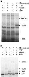
Fig 1.
Fig 1.
Affinity crosslinking between an activating region of Gal4 and the RNA polymerase II holoenzyme. (A) Purified native Gal4 (1–100) + (840–881) with a solvent-accessible cysteine at residue 856 was conjugated with 125I-APDP. The label-transfer crosslinking reaction with this modified protein was performed with the holoenzyme complex (lane 2), holoenzyme supplemented with TBP (lane 3), and holoenzyme and TBP in the presence of Gal80 (lane 4). The radiolabeled bands were resolved on an SDS/PAGE gel and visualized by phosphorimaging as described (10, 11). (B) Purified denatured Gal4 (1–100) + (840–881) was refolded and then conjugated to 125I-PEAS/AET. We confirmed the ability of this protein to bind DNA and activate transcription in vitro (data not shown). UV-induced crosslinking reactions were then performed in the presence of Gal80 (lane 2), TBP (lane 3), holoenzyme supplemented with TBP (lane 4), and holoenzyme, TBP, and Gal80 (lane 5).
Fig 2.
Fig 2.
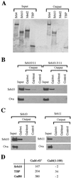
Gal4–SRB10 interaction studies in vitro. (A) In vitro-transcribed and -translated, 35S-labeled SRB4, SRB10, Med2, and TBP were separately incubated with the fusion protein GST-Gal4wt (840–874). The GST fusion and bound proteins were isolated by using glutathione-agarose beads. Ten percent of the input and the entire pellet (output) were subjected to SDS/PAGE and analyzed by autoradiography. In vitro-translated and 35S-labeled proteins were identified by their apparent molecular weights. (B) Recombinant SRB10/11 and SRB10–3/11 complex, purified from a baculoviral expression system, were separately incubated with GST-GAL4 (840–874) (GAL4wt) and with an otherwise identical protein bearing amino acid changes in the activating region (V864E and L868V) and called Gal4mut. This double mutation abolishes Gal4's ability to activate transcription. The GST control was included (Control), and ovalbumin (Ova) was added to each reaction mixture to serve as a control for specific precipitation. Bound proteins were subjected to SDS/PAGE as in A, and SRB10 and SRB10-3 were visualized by immunoblotting with an anti-SRB10 polyclonal antibody. (C) Insect cell extracts containing either SRB10 or SRB11 were incubated with proteins bearing GST fused to WT or mutant Gal4-activating region (840–874). GST pull-down and subsequent visualization of bound proteins were performed by using polyclonal antibodies against SRB10 and SRB11 as described in B. (D) SPR analysis was performed with purified recombinant SRB10/11 complex, TBP, and Gal80. In each case, ≈200 resonance response units of Gal4 (1–100) + rII′ or a fragment comprising Gal4 (1–100) was prebound to a DNA fragment bearing a Gal4 DNA binding site immobilized on the surface of a flow cell. Target proteins were passed over this Gal4–DNA complex, and the change in resonance response units (ΔRU) upon binding of target proteins to the respective Gal4 derivative is presented.
Fig 3.
Fig 3.
Gal4p interacts with SRB10 in vivo in the split-ubiquitin assay. Tenfold serial dilutions of yeast cells coexpressing Nub or Nub-Gal4 derivatives together with SRB10-Cub-Rura3 were spotted on a control plate lacking tryptophan and leucine (WL) or on plates additionally containing 5-FOA. Growth on either plate requires functions supplied by the plasmids, and the presence of FOA selects against Ura3.
Fig 4.
Fig 4.
Activating regions from different activators bind SRB10. In vitro-translated, 35S-labeled SRB10 was incubated with GST alone (control) or proteins comprising GST fused to one or another activating region as shown. The composition of each activating region is Gal4 (residues 840–874), AH (PGIELQELQELQALLQQQ), VP16 (residues 420–490), p53 (residues 1–97), Gcn4 no. 4 (107–144). For details, see ref. . Fractions (10%) of the input and the entire pellet (output) were analyzed as in Fig. 2A.

References

    1. Chi Y., Huddleston, M. J., Zhang, M., Young, R. A., Annan, R. S., Carr, S. A. & Deshaies, R. H. (2001) Genes Dev. 15, 1078-1092. - PMC - PubMed
    1. Hirst M., Kobor, M. S., Kuriakose, N., Greenblatt, J. & Sadowski, I. (1999) Mol. Cell 3, 673-678. - PubMed
    1. Vincent O., Kuchin, S., Hong, S. P., Townley, R., Vyas, V. K. & Carlson, M. (2001) Mol. Cell. Biol. 21, 5790-5796. - PMC - PubMed
    1. Sadowski I., Niedbala, D., Wood, K. & Ptashne, M. (1991) Proc. Natl. Acad. Sci. USA 88, 10510-10514. - PMC - PubMed
    1. Liao S. M., Zhang, J., Jeffery, D. A., Koleske, A. J., Thompson, C. M., Chao, D. M., Viljoen, M., van Vuuren, H. J. J. & Young, R. A. (1995) Nature 374, 193-196. - PubMed

Publication types

MeSH terms

LinkOut - more resources