. 2002 Nov 12;99(23):15124-9.
doi: 10.1073/pnas.232588699. Epub 2002 Nov 4.

Engineered herpes simplex virus 1 is dependent on IL13Ralpha 2 receptor for cell entry and independent of glycoprotein D receptor interaction

Affiliations

Engineered herpes simplex virus 1 is dependent on IL13Ralpha 2 receptor for cell entry and independent of glycoprotein D receptor interaction

Guoying Zhou et al. Proc Natl Acad Sci U S A. .

Abstract

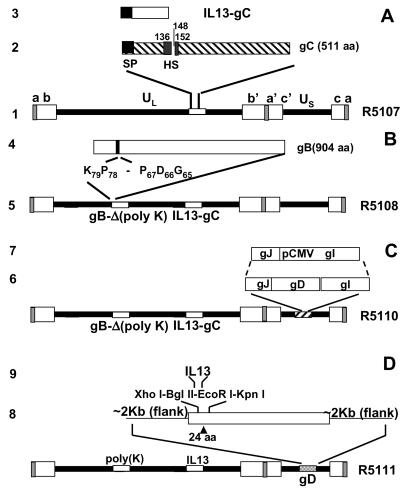
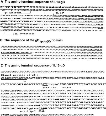
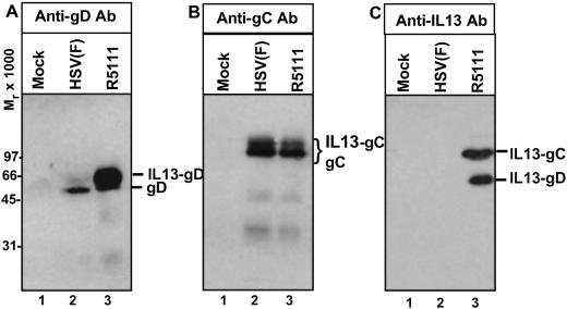
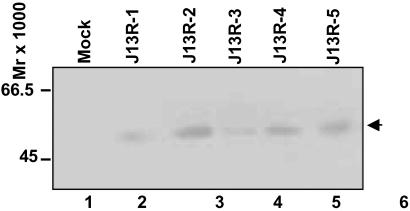
In the first stage of engineering a herpes simplex virus (HSV)-1 that specifically targets human malignant glioma cells, we constructed a recombinant virus designated R5111 in which we have ablated the binding sites for sulfated proteoglycans in glycoproteins B and C, replaced the amino-terminal 148 aa in glycoprotein C by IL-13 flanked at its amino terminus with a signal peptide, and inserted a second copy of IL-13 after the amino acid 24 of glycoprotein D. In the process, the binding site for HveA, a viral entry receptor, was disrupted. We have also transformed a cell line (J1.1) lacking HSV-1 receptors to express IL13Ralpha2 receptor (J13R cells). We report the following: the R5111 recombinant virus replicates as well as wild-type virus in a variety of cell lines including cell lines derived from brain tumors. R5111 failed to replicate in the parent J1.1 cell line but multiplied to titers similar to those obtained in other cell lines in the J13R cell line. On the basis of the evidence that R5111 can use the IL13Ralpha2 receptor for entry, we conclude that HSV-1 can use receptors other than HveA or nectins, provided it can bind to them. The domains of gD that interact with HveA and nectin receptors are independent of each other. Lastly, the fusogenic activities of the glycoproteins in the viral envelope are not dependent on a set of unique interactions between glycoprotein D and its receptor. The construction of R5111 opens the way for construction of viruses totally dependent on selected receptors for entry or imaging of targeted cells.


Figures

Fig 1.
Fig 1.
Schematic representation of the HSV-1 (F) genome and gene manipulations in glycoprotein C (gC), glycoprotein B (gB), and glycoprotein D (gD). Line 1, sequence arrangement of HSV-1 genome. The rectangular boxes represent the inverted repeat sequences ab and b′a′ flanking the unique long (UL) sequence and inverted repeat c′a′ and ca flanking the unique short (US) sequence. Line 2, sequence arrangement of the domains of the glycoprotein C, signal peptide (SP) domain, and heparan sulfate (HS)-binding domain of gC are highlighted. Line 3, human IL-13 with signal peptide that replaced the N-terminal segment of 148 aa of gC. Line 4, sequence arrangement of the polylysine domain of gB. Line 5, schematic representation of recombinant HSV-1(F) genome, in which the N-terminal domain of gC was replaced with IL-13, and the polylysine domain (from codon 68 to codon 77) of gB was deleted. Line 6, sequence arrangement of glycoprotein J (gJ), glycoprotein D (gD), and glycoprotein I (gI) in US. Line 7, replacement of gD with the immediately early promoter of cytomegalovirus to enable the expression of gI. Line 8, schematic representation of recombinant HSV-1(F) genome, in which the N-terminal domain of gC was replaced with IL-13, the polylysine domain of gB was deleted, and IL-13 was inserted after amino acid 24 of gD. Line 9, a polylinker XhoI-BglII-EcoRI-KpnI was inserted after amino acid 24 of gD, and then IL-13 was inserted into the XhoI and KpnI sites of gD.
Fig 2.
Fig 2.
Amino acid sequence alignment of IL-13–gC, IL-13–gD junction sequence and the HS-binding domain of gB. (A) The amino-terminal sequence of IL-13–gC chimeric protein. The sequences upstream and downstream of the HS-binding site portion are shown. IL-13 (27) was inserted between two restriction enzyme sites that are underlined. (B) The domain of the gB ORF from which the polylysine [poly(K)] sequence was deleted. The underlined sequences (codons 68–77) were not present in gB amplified from R5107. (C) The amino-terminal sequence of IL-13–gD. The first underlined sequence identifies the gD signal peptide. IL-13 (bracketed by arrows) was inserted between residues 24 and 25 (underlined) of gD, between the XhoI and KpnI restriction enzyme sites.
Fig 3.
Fig 3.
Verification of R5111 viral DNA by PCR. Photographs of electrophoretically separated PCR products amplified directly from the plaques picked from Vero (A) and HEp-2 cells (B). Viral DNA was extracted as described in Materials and Methods and subjected to PCR with IL-13 primers from IL-13 ORF and IL-13–gD primers, which bracketed IL-13 and the gD ectodomain.
Fig 4.
Fig 4.
Photograph of electrophoretically separated proteins from lysates of cells infected with R5111 reacted with antibody to gC, gD, or IL-13. HEp-2 grown in 25-cm2 flasks were exposed to 10 pfu of HSV-1 or R5111 per cell. The cells were harvested 24 h after infection, solubilized, subjected to electrophoresis in 10% denaturing polyacrylamide gels, electrically transferred onto a nitrocellulose sheet, and reacted with a monoclonal antibody against gD (A), gC (B), or IL-13 (C), respectively. The protein bands corresponding to the gC, IL-13–gC fusion protein, gD, and IL-13–gD fusion protein are indicated. IL-13–gC is expected to be the same size as the native gC.
Fig 5.
Fig 5.
HA-tagged IL13Rα2 expression from the individual clones of stable transfectants of the J1.1 cell line. The individual clones were amplified as detailed in Materials and Methods. The cells were harvested from 25-cm2 flasks, solubilized, and subjected to electrophoresis in 12% denaturing polyacrylamide gels, electrically transferred onto a nitrocellulose sheet, and reacted with a polyclonal antibody to HA tag.

References

    1. Davis F. G., Freels, S., Grutsch, J., Barlas, S. & Brem, S. (1998) J. Neurosurg. 88, 1-10. - PubMed
    1. Pyles R. B., Warnick, R. E., Chalk, C. L., Szanti, B. E. & Parysek, L. M. (1997) Hum. Gene Ther. 8, 533-544. - PubMed
    1. Rampling R., Cruickshank, G., Papanastassiou, V., Nicoll, J., Hadley, D., Brennan, D., Petty, R., MacLean, A., Harland, J., McKie, E., et al. (2000) Gene Ther. 7, 859-866. - PubMed
    1. McKie E. A., Brown, S. M., MacLean, A. R. & Graham, D. I. (1998) Neuropathol. Appl. Neurobiol. 24, 367-372. - PubMed
    1. Markert J. M., Medlock, M. D., Rabkin, S. D., Gillespie, G. Y., Todo, T., Hunter, W. D., Palmer, C. A., Feigenbaum, F., Tornatore, C., Tufaro, F. & Martuza, R. L. (2000) Gene Ther. 7, 867-874. - PubMed

Publication types