. 2002 Nov 12;99(23):14943-5.
doi: 10.1073/pnas.242594499. Epub 2002 Nov 4.

Retroviral delivery of small interfering RNA into primary cells

Affiliations

Retroviral delivery of small interfering RNA into primary cells

Gregory M Barton et al. Proc Natl Acad Sci U S A. .

Abstract

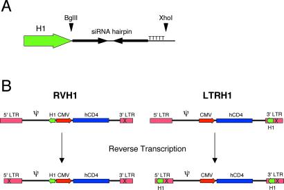
RNA interference is an evolutionarily conserved process in which recognition of double-stranded RNA ultimately leads to posttranscriptional suppression of gene expression. This suppression is mediated by short (21- to 22-nt) small interfering RNAs (siRNAs), which induce degradation of mRNA based on complementary base pairing. The silencing of gene expression by siRNAs is emerging rapidly as a powerful method for genetic analysis. Recently, several groups have reported systems designed to express siRNAs in mammalian cells through transfection of either oligonucleotides or plasmids encoding siRNAs. Because these systems rely on transfection for delivery, the cell types available for study are restricted generally to transformed cell lines. Here, we describe a retroviral system for delivery of siRNA into cells. The use of retroviral vectors can greatly expand the types of cells available for RNA interference analysis. Furthermore, we demonstrate that this retroviral system allows for stable inactivation of genes in primary cells.


Figures

Fig 1.
Fig 1.
(A) Schematic representation of the H1 promoter cassette. Unique restriction endonuclease sites allow cloning of hairpin-encoding oligonucleotides directly into the retroviral vectors. The five thymidines serve as a termination signal for RNA pol III. (B) Schematic representations of RVH1 and LTRH1 retroviral vectors. The vectors are shown above and the integrated proviruses are shown below. During reverse transcription, portions of the 3′ LTR serve as the template for generating the new 5′ LTR. The “X” in the 3′ LTR in the upper schematics represents deletion of the U3 region, which is copied into the 5′ LTR.
Fig 2.
Fig 2.
Infection of 293T cells with RVH1-p53 or LTRH1-p53 eliminates expression of p53. (A) CD4 expression on 293T cells infected with RVH1 vector, RVH1-p53, or LTRH1-p53 or mock-infected, as measured by flow cytometry. The percentage of infected cells 3 days postinfection is indicated. (B) 293T cells infected as in A were analyzed for p53 expression by Western blotting. Western blots are shown 3 and 6 days postinfection.
Fig 3.
Fig 3.
Multiple copies of the H1 cassette are necessary for complete silencing. (A) 293T cells were infected with serial dilutions of LTRH1-p53, and p53 expression was measured 3 days postinfection. The percentage of infected cells was determined by measuring CD4 expression by flow cytometry. The infection percentage and mean fluorescence intensity corresponding to each dilution are indicated. (B) 293T cells infected as described in A were analyzed for p53 expression by Western blot. (C) 293T cells infected as in A were analyzed for integration of the retroviral vector by Southern blot. A probe corresponding to the CMV promoter was used to detect the integrated viruses within genomic DNA from infected cells.
Fig 4.
Fig 4.
Retroviral delivery of siRNA into primary fibroblasts eliminates p53 expression. (A) FACS analysis of CD4 expression on primary human fibroblasts infected with LTRH1-p53 or mock-infected. (B) Primary human fibroblasts (AG01522 cells) were treated with 100 μM etoposide for the indicated times, after which the cells were harvested and p53 and p21 expression levels were determined by Western blot analysis. (C) Primary fibroblasts were infected with LTRH1-p53 virus as in A. Three days postinfection, cells were treated with 100 μM etoposide (etop). After 4 h of etoposide treatment, cells were harvested and expression levels of p53 and p21 were measured by Western blot analysis.

References

    1. Williams B. R. (1999) Oncogene 18, 6112-6120. - PubMed
    1. Sharp P. A. (2001) Genes Dev. 15, 485-490. - PubMed
    1. Fire A., Xu, S., Montgomery, M. K., Kostas, S. A., Driver, S. E. & Mello, C. C. (1998) Nature 391, 806-811. - PubMed
    1. Elbashir S. M., Lendeckel, W. & Tuschl, T. (2001) Genes Dev. 15, 188-200. - PMC - PubMed
    1. Elbashir S. M., Harborth, J., Lendeckel, W., Yalcin, A., Weber, K. & Tuschl, T. (2001) Nature 411, 494-498. - PubMed

Publication types

Substances