. 2002 Nov 12;99(23):14646-51.
doi: 10.1073/pnas.232299399. Epub 2002 Nov 4.

S-layer-streptavidin fusion proteins as template for nanopatterned molecular arrays

Affiliations

S-layer-streptavidin fusion proteins as template for nanopatterned molecular arrays

Dieter Moll et al. Proc Natl Acad Sci U S A. .

Abstract

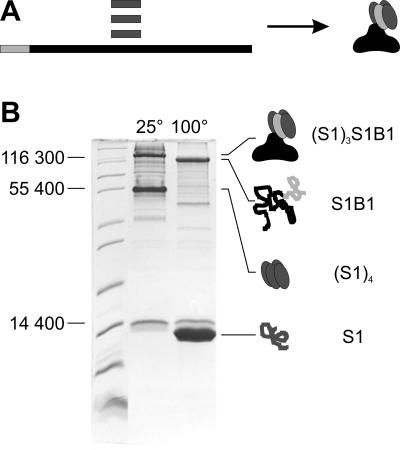
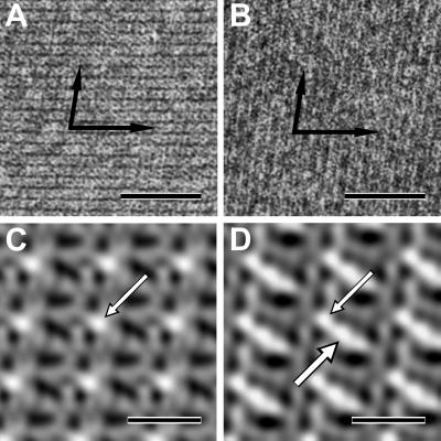
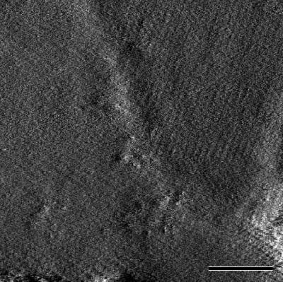
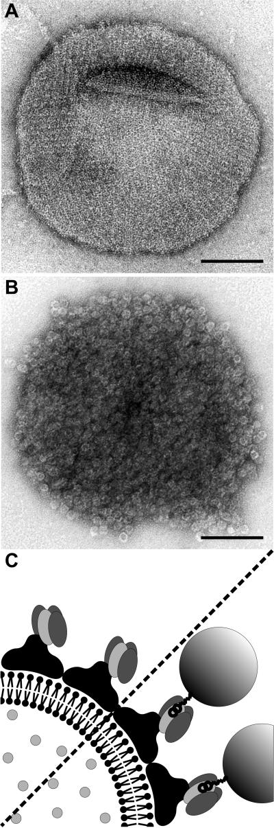
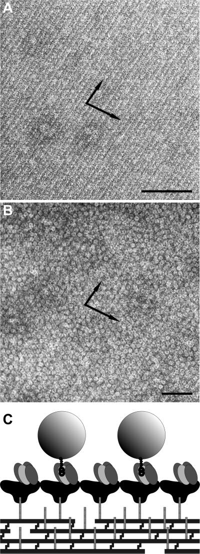
Biomolecular self-assembly can be used as a powerful tool for nanoscale engineering. In this paper, we describe the development of building blocks for nanobiotechnology, which are based on the fusion of streptavidin to a crystalline bacterial cell surface layer (S-layer) protein with the inherent ability to self-assemble into a monomolecular protein lattice. The fusion proteins and streptavidin were produced independently in Escherichia coli, isolated, and mixed to refold and purify heterotetramers of 1:3 stoichiometry. Self-assembled chimeric S-layers could be formed in suspension, on liposomes, on silicon wafers, and on accessory cell wall polymer containing cell wall fragments. The two-dimensional protein crystals displayed streptavidin in defined repetitive spacing, and they were capable of binding d-biotin and biotinylated proteins. Therefore, the chimeric S-layer can be used as a self-assembling nanopatterned molecular affinity matrix to arrange biotinylated compounds on a surface. In addition, it has application potential as a functional coat of liposomes.


Figures

Fig 1.
Fig 1.
Refolding of heterotetrameric fusion protein (S1)3S1B1 (A) and confirmation by SDS/PAGE analysis (B). (A) Core streptavidin (S1, dark gray) and fusion protein S1B1 (streptavidin moiety S1, light gray; SbsB moiety B1, black) were produced in E. coli and isolated independently, mixed, and refolded to heterotetramers (S1)3S1B1. (B) For SDS/PAGE analysis, samples were recovered from a refolding batch, and SDS was added to a concentration of 5%. Without boiling of the sample (lane 25°C), streptavidin–SbsB heterotetramers (S1)3S1B1 (144,973 Da) and core streptavidin homotetramers (S1)4 (49,840 Da) migrated at apparent molecular masses of 125,000 and 55,000 Da, respectively, on an SDS/15% PAGE gel. After boiling (lane 100°C), the tetramers of both the fusion protein and core streptavidin were denatured to monomers (S1B1, 107,593 Da; S1, 12,460 Da).
Fig 2.
Fig 2.
Self-assembly products and digital image reconstructions of SbsB (see A and C for comparison) and fusion protein (S1)3S1B1 (B and D). (A and B) The crystalline sheets were formed in suspension and negatively stained with uranyl acetate for TEM. The arrows indicate the base vectors of the oblique p1 lattice. (Bars = 50 nm.) (C and D) The digital image reconstructions were made by Fourier processing of electron micrographs (not identical to the ones shown in A and B). The region of highest protein mass in the SbsB lattice is the SLH-domain (C, arrow). In the lattice of the fusion protein, streptavidin showed up as additional protein mass (D, thick arrow) attached to the SLH domain. (Bars = 10 nm.)
Fig 3.
Fig 3.
Fusion protein (S1)3S1B1 crystallized on silicon wafers. The atomic force microscopy deflection image was recorded in contact mode under 100 mM NaCl; z-range = 0.6 nm. (Bar = 100 nm.)
Fig 4.
Fig 4.
Liposomes carrying a chimeric S-layer formed by the fusion protein (S1)3S1B1 (A) were capable of binding biotinylated ferritin (B). Preparations were negatively stained with uranyl acetate for TEM. (Bars = 100 nm.) (C) The cartoon shows the orientation of (S1)3S1B1 on liposomes with the streptavidin-carrying inner face of the S-layer exposed.
Fig 5.
Fig 5.
Cell wall fragments carrying a chimeric S-layer formed by the fusion protein BS1(S1)3 (A) were capable of binding biotinylated ferritin (B). (A) Self-assembly was enabled by the specific interaction between an accessory cell wall polymer that is part of the cell wall of G. stearothermophilus PV72/p2 and the SLH domain of the fusion protein. (B) Bound biotinylated ferritin reflected the underlying S-layer lattice. The preparations were negatively stained with uranyl acetate for TEM. The arrows indicate the base vectors of the oblique p1 lattice. (Bars = 100 nm.) (C) The cartoon shows the orientation of BS1(S1)3 after SLH-enabled self-assembly with the streptavidin-carrying outer face of the S-layer exposed.

References

    1. Winfree E., Liu, F., Wenzler, L. A. & Seeman, N. C. (1998) Nature 394, 539-544. - PubMed
    1. Seeman N. C. (1999) Trends Biotechnol. 17, 437-443. - PubMed
    1. Niemeyer C. M. (2000) Curr. Opin. Chem. Biol. 4, 609-618. - PubMed
    1. Padilla J. E., Colovos, C. & Yeates, T. O. (2001) Proc. Natl. Acad. Sci. USA 98, 2217-2221. - PMC - PubMed
    1. Sleytr U. B., Sára, M. & Pum, D. (2000) in Supramolecular Polymers, ed. Ciferri, A. (Dekker, New York), pp. 177–213.

Publication types