

Motion perception of saccade-induced retinal translation
- PMID: 12417765
- PMCID: PMC137560
- DOI: 10.1073/pnas.232377199
Motion perception of saccade-induced retinal translation
Abstract
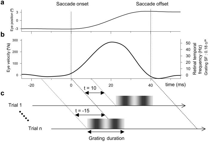
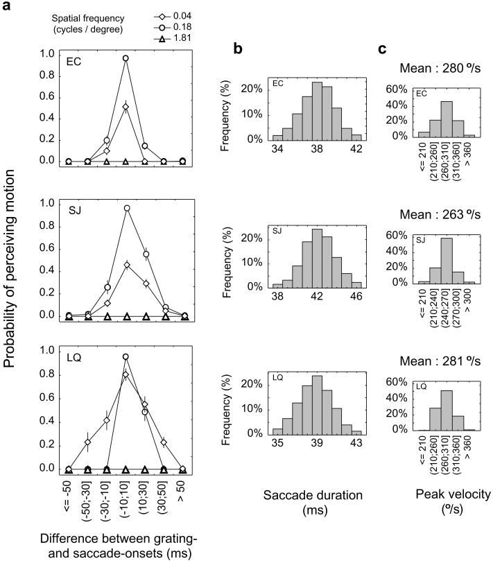
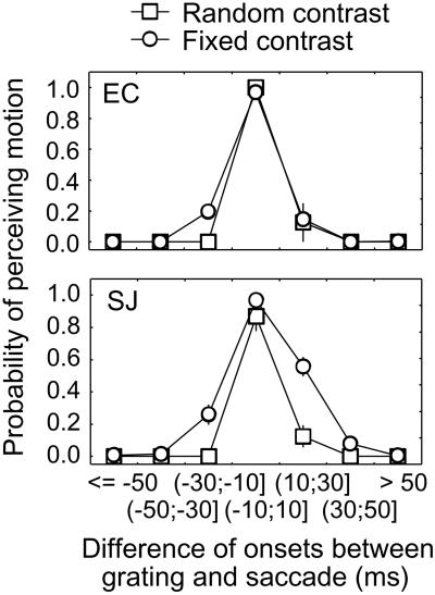
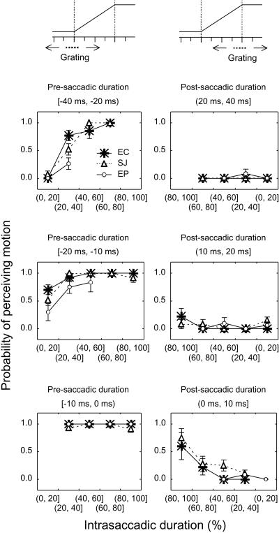
Active visual perception relies on the ability to interpret correctly retinal motion signals induced either by moving objects viewed with static eyes or by stationary objects viewed with moving eyes. A motionless environment is not normally perceived as moving during saccadic eye movements. It is commonly believed that this phenomenon involves central oculomotor signals that inhibit intrasaccadic visual motion processing. The keystone of this extraretinal theory relies on experimental reports showing that physically stationary scenes displayed only during saccades, thus producing high retinal velocities, are never perceived as moving but appear as static blurred images. We, however, provide evidence that stimuli optimized for high-speed motion detection elicit clear motion perception against saccade direction, thus making the search for extraretinal suppression superfluous. The data indicate that visual motion is the main cue used by observers to perform the task independently of other perceptual factors covarying with intrasaccadic stimulation. By using stimuli of different durations, we show that the probability of perceiving the stimulus as static, rather than moving, increases when the intrasaccadic stimulation is preceded or followed by a significant extrasaccadic stimulation. We suggest that intrasaccadic motion perception is accomplished by motion-selective magnocellular neurons through temporal integration of rapidly increasing retinal velocities. The functional mechanism that usually prevents this intrasaccadic activity from being perceived seems to rely on temporal masking effects induced by the static retinal images present before and/or after the saccade.
Figures
References
-
- Matin E., Clymer, A. B. & Matin, L. (1972) Science 178, 179-182. - PubMed
-
- Campbell F. W. & Wurtz, R. H. (1978) Vision Res. 18, 1297-1303. - PubMed
-
- Mitrani L., Mateeff, S. & Yakimoff, N. (1970) Vision Res. 10, 405-409. - PubMed
-
- Burr D. C. & Ross, J. (1982) Vision Res. 22, 479-484. - PubMed
-
- Ross J., Burr, D. & Morrone, C. (1996) Behav. Brain Res. 80, 1-8. - PubMed
MeSH terms
LinkOut - more resources
Full Text Sources
