. 2002 Nov 12;99(23):15006-11.
doi: 10.1073/pnas.232058599. Epub 2002 Nov 5.

Human CD8+ T cells do not require the polarization of lipid rafts for activation and proliferation

Affiliations

Human CD8+ T cells do not require the polarization of lipid rafts for activation and proliferation

Birgit Kovacs et al. Proc Natl Acad Sci U S A. .

Abstract

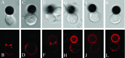
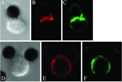
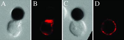
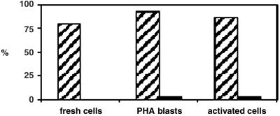
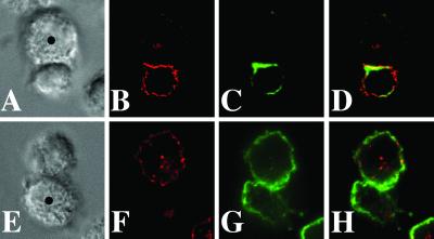
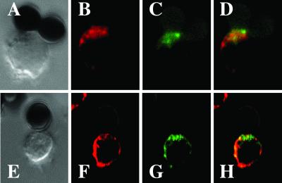
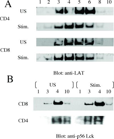
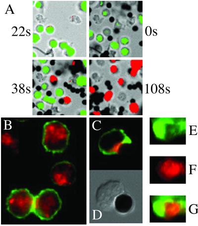
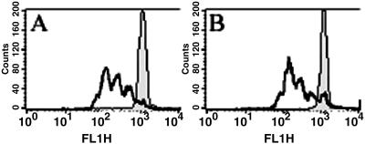
Lipid rafts are important signaling platforms in T cells. Little is known about their properties in human CD8(+) T cells. We studied polarization of lipid rafts by digital immunofluorescence microscopy in primary human T cells, using beads coated with anti-CD3 and anti-CD28 mAbs (CD3/28 beads). Unlike CD4(+) T cells, CD8(+) T cells did not polarize lipid rafts when stimulated with CD3/28 beads, when the anti-CD28 antibody was substituted with B7.2Ig, or if an anti-CD8 antibody was added to the CD3/28 beads. This phenomenon was also observed in human antigen-specific CD8(+) T cells. On stimulation with CD3/28 beads, the T cell antigen receptor clustered at the cell/bead contact area in both CD4(+) and CD8(+) T cells. Examination of lipid rafts isolated by sucrose density gradient centrifugation revealed the constitutive expression of p(56)Lck in the raft fractions of unstimulated CD8(+) T cells, whereas p(56)Lck was recruited to the raft fraction of CD4(+) T cells only after stimulation with CD3/28 beads. Stimulation with CD3/28 beads induced marked calcium flux, recruitment of PKC-theta and F-actin to the cell/bead contact site, and similar proliferation patterns in CD4(+) and CD8(+) T cells. Thus, polarization of lipid rafts is not essential for early signal transduction events or proliferation of human CD8(+) lymphocytes. It is possible that the lower stringency of CD8(+) T cell activation obviates a requirement for raft polarization.


Figures

Fig 1.
Fig 1.
Primary CD8+ T cells do not polarize lipid rafts at the site of TCR engagement. CD4+ (B, F, and J) or CD8+ (D, H, and L) T cells were incubated with CD3/28 beads for 30 min and stained with anti-GM1 antiserum. The corresponding Nomarski images are shown in the top row. In EH, cells were incubated with beads coated with a ratio of anti-CD3 to anti-CD28 of 15:85. In IL, the ratio was 5:95. The data are representative of three experiments with different human donors.
Fig 2.
Fig 2.
Long-term activated CD8+ T cells do not polarize lipid rafts. CD4+ (AC) or CD8+ (DF) T cells that had been activated with PHA and IL-2 for 7 days were incubated with CD3/28 beads for 20 min and stained with anti-GM1 antiserum (red, B and E) and cholera toxin B-FITC (green, C and F). Corresponding Nomarski images are shown in A and D. Data are representative of three different experiments.
Fig 3.
Fig 3.
Replacement of the CD28 antibody with B7.2Ig on the beads does not reconstitute the polarization of lipid rafts in CD8+ T cells. CD4+ (A and B) or CD8+ (C and D) T cells that had been activated with PHA for 2 days were incubated with CD3/B7.2Ig beads for 30 min and stained with anti-GM1 antiserum. Corresponding Nomarski images are shown in A and C. The pictures are representative of four different experiments.
Fig 4.
Fig 4.
Quantitative analysis of cell/bead conjugates showing aggregation of GM1. For both CD4+ and CD8+ cells, 30 conjugates each were scored in primary cells, PHA blasts, and long-term activated cells after stimulation with CD3/28 beads for 30 min. The numbers indicate the percentage of conjugates scored that exhibit aggregation of GM1 at the cell/bead contact site. Data are compiled from three different experiments for each cell activation condition.
Fig 5.
Fig 5.
Human antigen-specific CD8+ T cells do not polarize lipid rafts to the APC-T cell contact area, but recruit F-actin to the immunological synapse. FLU-specific CD8+ T cells were prelabeled with CTB-Bio and incubated with EBV-transformed B cells as APCs (•) for 30 min. In AD, APCs were pulsed with FLU peptide overnight, and in EH, APCs remained nonpulsed. CTB stain is shown in red (B and F) and F-actin stain is shown in green (C and G). (D and H) The overlay of both stains. Corresponding Nomarski images are shown in A and E. The pictures are representative of two different experiments. Ten conjugates were examined for each condition. We observed recruitment of F-actin to the immunological synapse in 8 of 10 conjugates.
Fig 6.
Fig 6.
Primary CD8+ T cells recruit the TCR to the cell–bead contact area. CD4+ (BD) or CD8+ (FH) T cells were incubated with CD3/28 beads for 30 min and stained with anti-GM1 antiserum (red, B and F) or anti-TCR antibody (green, C and G). (D and H) The overlay of both stains. The corresponding Nomarski images are shown in A and E. The pictures are representative of three different experiments.
Fig 7.
Fig 7.
Identification of raft fractions and targeting of p56Lck to lipid rafts in human CD4+ and CD8+ T cells. Primary human T cells were either unstimulated (US) or stimulated with CD3/28 beads for 10 min, followed by lysis in Mes lysis buffer plus protease and phosphatase inhibitors. Lysates were subjected to sucrose density gradient centrifugation for lipid raft purification. Numbers denote gradient fractions. The gradient fractions were separated by SDS/PAGE , followed by detection of LAT (A) and Lck (B) with immunoblot analysis.
Fig 8.
Fig 8.
Early signal transduction events are intact in CD8+ T cells activated with CD3/28 beads. (A) CD8+ T cells show intracellular calcium flux on stimulation with CD3/28 beads. Each picture shows the composition of a bright-field image overlaid with a transparent color scale of the ratio of fluorescence emissions at 340 and 380 nm of fura-2. The ratio correlates with intracellular calcium concentration: green, resting (low); red, high intracellular calcium concentration. The picture taken at t = −22 s shows CD8+ T cells in the resting state. At t = 0 s, the CD3/28 beads were added. At 108 s, ionomycin was added as a positive control. These data are representative of three experiments. (BG) PKC-θ and F-actin are recruited to the cell/bead contact site in CD8+ T cells. In resting T cells (B), PKC-θ (in red) is distributed in the cytoplasm and F-actin (in green) is distributed evenly in the plasma membrane. After activation of the CD8+ T cells for 30 min with CD3/28 beads, PKC-θ and F-actin are recruited to the cell/bead contact site (C). (D) The Nomarski image of the cell/bead conjugate shown in C. Three-dimensional volume rendering shows distribution of F-actin in the peripheral SMAC (E) and PKC-θ in the central SMAC (F). (G) The overlay of pictures D and E. The data are representative of three different experiments.
Fig 9.
Fig 9.
CD8+ and CD4+ T cells show a similar proliferation profile after activation with CD3/28 beads. CD4+ (A) and CD8+ (B) T cells were labeled with CFSE, activated with CD3/28 beads for 3 days, and analyzed by flow cytometry. Unstimulated cells that were labeled with CFSE were used as controls. Data are representative of two different experiments.

References

    1. Monks C. R. F., Freiberg, B. A., Kupfer, H., Sciaky, N. & Kupfer, A. (1998) Nature 395, 82-86. - PubMed
    1. Cherukuri A., Dykstra, M. & Pierce, S. K. (2000) Immunity 14, 657-660. - PubMed
    1. Viola A., Schroeder, S., Sakakibara, Y. & Lanzavecchia, A. (1999) Science 283, 680-682. - PubMed
    1. Xavier R., Brennan, T., Li, Q., McCormack, C. & Seed, B. (1998) Immunity 8, 723-732. - PubMed
    1. Bi K., Tanaka, Y., Coudronniere, N., Sugie, K., Hong, S., van Stipdonk, M. J. B. & Altman, A. (2001) Nat. Immunol. 2, 556-562. - PubMed

Publication types

MeSH terms