. 2002 Nov;107(3):350-7.
doi: 10.1046/j.1365-2567.2002.01511.x.

Reactivation of latent tuberculosis by an inhibitor of inducible nitric oxide synthase in an aerosol murine model

Affiliations

Reactivation of latent tuberculosis by an inhibitor of inducible nitric oxide synthase in an aerosol murine model

Tania Botha et al. Immunology. 2002 Nov.

Abstract

Exposure to Mycobacterium tuberculosis results in clinical tuberculosis only in a small percentage of healthy individuals. In most instances the bacilli are controlled by the immune system and survive in a latent state within granuloma. Immunosuppression, however, may result in reactivation of infection, resulting in clinical disease. Using a low-dose aerosol infection (30 colony-forming units) in mice, we describe a short-duration model for studying spontaneous and drug-induced reactivation of anti-tuberculous drug-treated, latent tuberculosis infection. Although a 4-week treatment with rifampicin and isoniazid reduced the number of bacilli to undetectable levels, the infection spontaneously reactivated following therapy. By contrast, an 8-week treatment period induced a state of latent infection, requiring immunosuppression to reactivate infection. Finally, a 12-week treatment period eliminated the bacilli completely and aminoguanidine did not induce reactivation of infection. In view of the fact that therapy in the selected protocol reduces the mycobacterial load to undetectable levels, the data suggest that an 8-week treatment period is necessary and sufficient to mount protective immunity in mice.


Figures

Figure 1
Figure 1
Schematic diagram of the experimental design for establishing the optimal duration of rifampicin and isoniazid (RMP-INH) treatment in mice to achieve latency of tuberculosis. Unfilled bars depict RMP-INH therapy, while black bars indicate periods of aminoguanidine administration. CFU, colony-forming units.
Figure 2
Figure 2
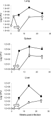
Model A. Bacterial burden of C57BL/6 mice treated with rifampicin and isoniazid (RMP-INH) for 4 weeks after aerogenic infection with 30 colony-forming units (CFU) of Mycobacterium tuberculosis H37Rv. Two weeks postinfection, one group of mice (○) received 0·1 g/l RMP-INH, delivered in drinking water ad libitum (indicated by arrows) for a period of 4 weeks, whereas infected control mice (•) received plain drinking water ad libitum. At specific time-intervals up to 24 weeks postinfection, the number of viable bacilli were determined in the lung, spleen and liver by plating organ homogenates and enumerating CFU. No viable bacilli were detected in organs of mice upon cessation of anti-tuberculous therapy; however, mice spontaneously reactivated and bacilli were cultured 8 weeks later (open circles). Both infected controls and reactivated mice controlled the infection (100% survival). Results are expressed as means ± SEM and are representative of three independent experiments with three to four mice per group per time-point.
Figure 3
Figure 3
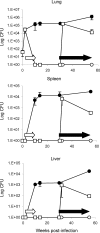
Model B. Bacterial burden of C57BL/6 mice treated with rifampicin and isoniazid (RMP-INH) for 8 weeks after aerogenic infection with 30 colony-forming units (CFU) of Mycobacterium tuberculosis H37Rv. Two weeks postinfection, two groups of mice (○ and □) received 0·1 g/l RMP-INH, delivered in drinking water ad libitum (open arrows) for a period of 8 weeks, whereas the control group (•) received plain drinking water ad libitum. At specific time-points up to 55 weeks postinfection, the number of viable bacilli in the lung, spleen and liver were determined by plating organ homogenates and enumerating CFU. No viable bacilli were detected in the organs of mice upon completion of chemotherapy. After administration of 2·5% aminoguanidine in drinking water containing 10% glucose (solid arrows) from weeks 30–55, reactivated infection was observed in 67% of mice (□, representative of reactivated mice only), although they survived (100% survival) and seemed to control the infection. The group of mice with latent infection (○) remained culture negative, with no viable CFU in organs detected for the duration of the experiment (55 weeks). Infected control mice (•) showed a rapid initial increase in the CFU in lung, liver and spleen, although these mice contained the infection successfully for the duration of the experiment (100% survival). Results are expressed as means ± SEM and are representative of three independent experiments with three to four mice per group per time-point.
Figure 4
Figure 4
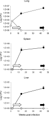
Model C. Bacterial burden of C57BL/6 mice treated with rifampicin and isoniazid (RMP-INH) for 12 weeks after aerogenic infection with 30 colony-forming units (CFU) of Mycobacterium tuberculosis H37Rv. Two weeks postinfection, one group of mice (○) received 0·1 g/l RMP-INH, delivered in drinking water ad libitum (open arrows) for a period of 12 weeks, whereas the control group (•) received plain drinking water ad libitum. At specific time-points up to 44 weeks postinfection, the number of viable bacilli in the lung, spleen and liver were determined by plating organ homogenates and enumerating CFU. No viable bacilli were detected in the organs of mice after completion of chemotherapy. Mice treated with RMP-INH for 12 weeks and subsequently with aminoguanidine for 4 weeks (solid arrows) failed to reactivate infection. Results are expressed as means ± SEM and are representative of three independent experiments with three to four mice per group per time-point.
Figure 5
Figure 5
Morphology of the lungs of mice during active, latent and reactivated tuberculosis infection. Lungs were examined at 2 weeks (a), 10 weeks (b) and (c), and 55 weeks (d)–(f), postinfection. Panel (a) represents a C57BL/6 mouse 2 weeks postinfection, prior to chemotherapy; panels (b) and (c) 10 weeks postinfection – an infected control (b) and a latently infected mouse after 8 weeks of rifampicin and isoniazid (RMP-INH) chemotherapy (c). Panels (d), (e) and (f) represent mice 55 weeks postinfection – an infected control [(d) and (f)] and a mouse reactivated after aminoguanidine administration (e). Sections were stained with haematoxylin and eosin [(a) to (e)] or with Ziehl–Neelsen (f). Magnification: panels (a)–(e) × 100; panel (f) × 400.
Figure 6
Figure 6
Inducible nitric oxide synthase (iNOS) expression in lung tissue in mice during active, latent and reactivated tuberculosis infection. Mice were aerogenically infected with 30 colony-forming units (CFU) of Mycobacterium tuberculosis H37Rv, after which a group was treated with rifampicin and isoniazid (RMP-INH) for 8 weeks to induce latency of infection. Lung tissue of C57BL/6 mice was examined at 32 weeks postinfection. No iNOS staining was detected in mice during latent infection (a), whereas intense iNOS expression was observed in infected control mice (b). An augmented iNOS staining pattern was observed in mice 2 weeks after aminoguanidine-induced reactivation (c). Panel (d) shows a granuloma in the liver of an infected control mouse to illustrate the origin of iNOS protein from activated macrophages. Magnification: panels (a), (b) and (c) × 100; (d) × 400.
Figure 7
Figure 7
Comparison of bacterial burden in lung, spleen and liver of mice following reactivation versus primary infection. C57BL/6 mice were infected with 30 colony-forming units (CFU) of Mycobacterium tuberculosis H37Rv and the number of viable CFU was determined 2 weeks postinfection (solid bars). Mice were then treated with rifampicin and isoniazid (RMP-INH) for 8 weeks to achieve latency (with no viable bacilli detected in all organs). After 20 weeks in this latent state, a group of mice received 2·5% aminoguanidine containing 10% glucose in drinking water ad libitum to induce reactivation. The increases in bacterial count following reactivated infection are shown 2 weeks after aminoguanidine administration (open bars). Results are expressed as means ± SEM and are representative of three independent experiments with four mice per group per time-point.

Similar articles

Cited by

References

    1. Sudre P, Ten Dam G, Kochi A. Tuberculosis: a global overview of the situation today. Bull WHO. 1992;70:149–59. - PMC - PubMed
    1. Kochi A. The global tuberculosis situation and the new control strategy of the World Health Organization. Tubercle. 1991;72:1–6. - PubMed
    1. Stead WW. Pathogenesis of a first episode of chronic pulmonary tuberculosis in man: recrudescence of residuals of the primary infection or exogenous reinfection? Am Rev Resp Dis. 1967;95:729–45. - PubMed
    1. Raviglione MC, Snider DE, Kochi A. Global epidemiology of tuberculosis: morbidity and mortality of a global epidemic. JAMA. 1995;273:220–6. - PubMed
    1. Antonucci G, Girardi E, Raviglione MC, Ippolito G. Risk factors for tuberculosis in HIV-infected persons: a prospective cohort study. JAMA. 1995;274:143–8. - PubMed

Publication types

MeSH terms