. 2002 Dec;71(6):1285-95.
doi: 10.1086/344515. Epub 2002 Nov 6.

Structural and functional characterization of factor H mutations associated with atypical hemolytic uremic syndrome

Affiliations

Structural and functional characterization of factor H mutations associated with atypical hemolytic uremic syndrome

Pilar Sánchez-Corral et al. Am J Hum Genet. 2002 Dec.

Abstract

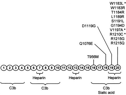
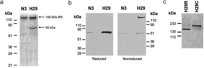
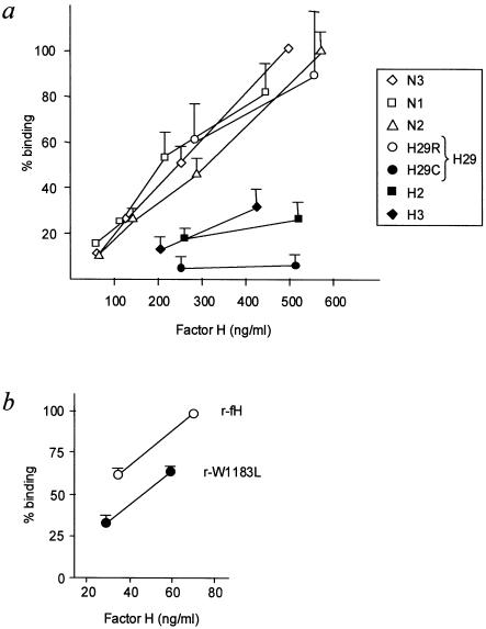
Genetic studies have demonstrated the involvement of the complement regulator factor H in nondiarrheal, nonverocytotoxin (i.e., atypical) cases of hemolytic uremic syndrome. Different factor H mutations have been identified in 10%-30% of patients with atypical hemolytic uremic syndrome (aHUS), and most of these mutations alter single amino acids in the C-terminal region of factor H. Although these mutations are considered to be responsible for the disease, the precise role that factor H plays in the pathogenesis of aHUS is unknown. We report here the structural and functional characterization of three different factor H proteins purified from the plasma of patients with aHUS who carry the factor H mutations W1183L, V1197A, or R1210C. Structural anomalies in factor H were found only in R1210C carriers; these individuals show, in their plasma, a characteristic high-molecular-weight factor H protein that results from the covalent interaction between factor H and human serum albumin. Most important, all three aHUS-associated factor H proteins have a normal cofactor activity in the proteolysis of fluid-phase C3b by factor I but show very low binding to surface-bound C3b. This functional impairment was also demonstrated in recombinant mutant factor H proteins expressed in COS7 cells. These data support the hypothesis that patients with aHUS carry a specific dysfunction in the protection of cellular surfaces from complement activation, offering new possibilities to improve diagnosis and develop appropriate therapies.


Figures

Figure  1
Figure 1
Functional domains and mutations in the factor H molecule, showing a diagram of the structure of human factor H with the 20 SCRs. Functional domains are indicated schematically. The location of the missense mutations thus far characterized in patients with aHUS (Warwicker et al. ; Caprioli et al. ; Pérez-Caballero et al. ; Richards et al. ; Perkins and Goodship ; Remuzzi et al. 2002b) is indicated, to illustrate that they are clustered in a specific factor H region that has been involved in the control of C3b deposited on surfaces. The mutations analyzed in the present article are indicated by asterisks.
Figure  2
Figure 2
Electrophoretic analysis of control and aHUS-associated factor H proteins. All samples included in the present analyses were run nonreduced in 8% polyacrylamide gels in the presence of SDS. a, 8% polyacrylamide gel stained with 1% Coomassie, to illustrate that no differences were observed between the factor H proteins purified from control individuals N1 and N2 (1 μg) and patients HUS2 and HUS3 (2 μg). b, 8% polyacrylamide gel stained with 1% Coomassie, showing that two protein bands, of 210 kDa and 155 kDa (factor H), were copurified from patient HUS29. c, Western blot using the 35H9 monoclonal anti–factor H antibody, to illustrate that the two protein bands purified from patient HUS29 are recognized as factor H. Factor H purified from control individual N3 was used as a reference of wild-type factor H in this experiment.
Figure  3
Figure 3
Segregation of the R1210C mutation in family HUS29. a, Pedigree of family HUS29, illustrating the segregation of the R1210C mutation. Levels of factor H in plasma are indicated for each member of the family. Nonaffected R1210C carriers are shown in gray. Seven SNPs in the factor H gene, including one in the promoter region (−331T/C), were analyzed to determine the segregation of the maternal and paternal factor H alleles in the patient and her sister, but the mother was homozygous for the seven SNPs. b, Western blot analysis of 1 μl of plasma from each HUS29 family member by using the anti–human factor H monoclonal antibody 35H9. Plasma samples are not reduced. The positions of the two factor H bands are indicated with arrows. Notice the decreased levels of both factor H alleles in the patient (IV-1).
Figure  4
Figure 4
Characterization of the high-molecular-weight factor H associated with the R1210C mutation. a, 10% SDS-PAGE of the factor H (fH) proteins purified from patient HUS29 and control individual N3. Samples were reduced with 10% β-mercaptoethanol. A gel stained with 1% Coomassie is shown. b, Western blot analyses of reduced and nonreduced factor H proteins purified from patient HUS29 and control individual N3, using a rabbit anti-HSA polyclonal antibody. c, SDS-PAGE analysis of the wild-type (H29R; flow through) and mutated (H29C; retained) factor H proteins purified from patient HUS29, using anti-HSA affinity chromatography. Samples were not reduced. A silver-stained 10% polyacrylamide gel is shown.
Figure  5
Figure 5
Analysis of the cofactor activity of factor H in the proteolysis of fluid-phase C3b by factor I. a, Analysis by 8% SDS-PAGE under reducing conditions of the proteolysis of C3b by factor I in the presence of factor H. Factor H samples include purified factor H from two control individuals (N1 and N2), purified factor H from patients with aHUS (HUS2 [H2] and HUS3 [H3]), purified factor H corresponding to the wild-type (H29R) and mutated (H29C) alleles of patient HUS29, and purified recombinant wild-type factor H (r-factor H) and W1183L mutant factor H (r-W1183L) expressed in COS7 cells. Each panel includes three different concentrations of factor H. Identification of the C3b fragments is depicted in the panel corresponding to N2. The cofactor activity of factor H results in cleavage of the α′ chain of C3b in the fragments α65 and α45/α43. The top left panel corresponds to the control lanes of C3b alone and C3b plus factor I in the absence of factor H. b, Densitometric analysis of the proteolysis of C3b by factor I in the presence of factor H. Stained gels were scanned in a GS-800 Calibrated Densitometer (Bio-Rad), and the bands were quantified with ImageQuant, version 3.3 (Molecular Dynamics). The percentage of the C3b cleavage was determined as the ratio of the values corresponding to the α65 and the α′ bands. The code for the samples is indicated at bottom.
Figure  6
Figure 6
Binding of factor H to surface-bound C3b. a, Analysis of factor H purified from plasma. Included were purified factor H from three control individuals (N1, N2, and N3), purified factor H from patients with aHUS (HUS2 [H2] and HUS3 [H3]), and purified factor H corresponding to the wild-type (H29R) and mutated (H29C) factor H alleles of patient HUS29. In the present analyses, the concentration of factor H ranged between 50 and 500 ng/ml. The results are expressed as the percentage of the binding observed for factor H from the control individual N3 at 500 ng/ml. Values are the mean ± SD of at least three independent experiments. b, Analysis of recombinant factor H proteins, showing the binding of purified recombinant wild-type factor H (r-fH) and W1183L mutant factor H (r-W1183L). Two different concentrations of recombinant factor H were used. The results are expressed as the percentage of the binding observed for recombinant wild-type factor H (r-fH) at the highest concentration. Values are the mean ± SD of at least three independent experiments.

References

Electronic-Database Information

    1. Entrez-Nucleotide, http://www.ncbi.nlm.nih.gov/entrez/query.fcgi?db=Nucleotide (for factor H cDNA sequence [accession number Y00716])
    1. Online Mendelian Inheritance in Man (OMIM), http://www.ncbi.nlm.nih.gov/Omim/ (for autosomal dominant [MIM 134370] and recessive [MIM 235400] HUS)

References

    1. Blackmore TK, Hellwage J, Sadlon TA, Higgs N, Zipfel PF, Ward HM, Gordon DL (1998) Identification of the second heparin-binding domain in human complement factor H. J Immunol 160:3342–3348 - PubMed
    1. Blackmore TK, Sadlon TA, Ward HM, Lublin DM, Gordon DL (1996) Identification of a heparin binding domain in the seventh short consensus repeat of complement factor H. J Immunol 157:5422–5427 - PubMed
    1. Buddles MR, Donne RL, Richards A, Goodship J, Goodship TH (2000) Complement factor H gene mutation associated with autosomal recessive atypical hemolytic uremic syndrome. Am J Hum Genet 66:1721–1722 - PMC - PubMed
    1. Caprioli J, Bettinaglio P, Zipfel PF, Amadei B, Daina E, Gamba S, Skerka C, Marziliano N, Remuzzi G, Noris M (2001) The molecular basis of familial hemolytic uremic syndrome: mutation analysis of factor H gene reveals a hot spot in short consensus repeat 20. J Am Soc Nephrol 12:297–307 - PubMed
    1. Fujita T, Nussenzweig V (1979) The role of C4-binding protein and β1H in proteolysis of C4b and C3b. J Exp Med 150:267–276 - PMC - PubMed

Publication types

MeSH terms

Associated data

LinkOut - more resources