Comparative Study
. 2002 Nov 15;21(22):6114-24.
doi: 10.1093/emboj/cdf608.

Structural basis for the Golgi membrane recruitment of Sly1p by Sed5p

Affiliations
Comparative Study

Structural basis for the Golgi membrane recruitment of Sly1p by Sed5p

Andreas Bracher et al. EMBO J. .

Abstract

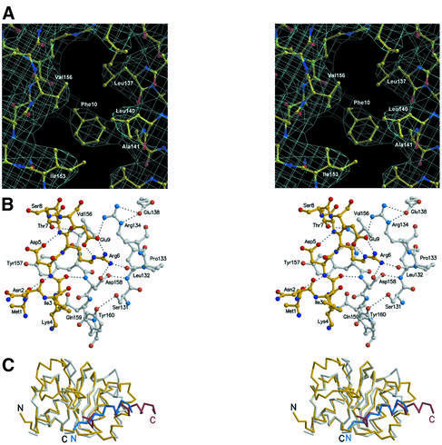
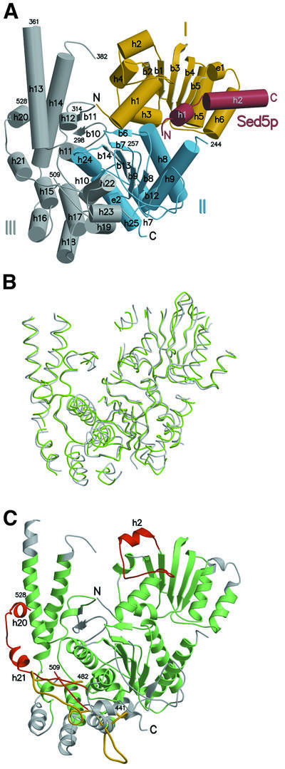
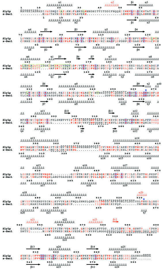
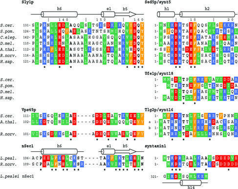
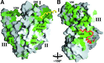
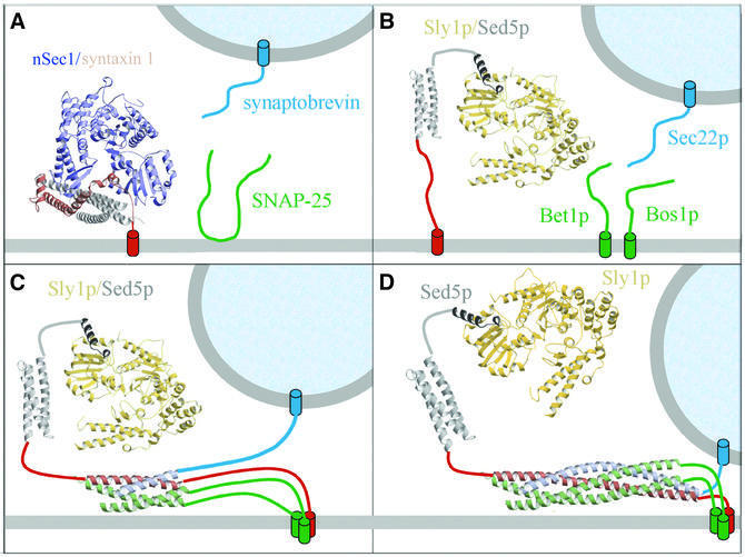
Cytosolic Sec1/munc18-like proteins (SM proteins) are recruited to membrane fusion sites by interaction with syntaxin-type SNARE proteins, constituting indispensable positive regulators of intracellular membrane fusion. Here we present the crystal structure of the yeast SM protein Sly1p in complex with a short N-terminal peptide derived from the Golgi-resident syntaxin Sed5p. Sly1p folds, similarly to neuronal Sec1, into a three-domain arch-shaped assembly, and Sed5p interacts in a helical conformation predominantly with domain I of Sly1p on the opposite site of the nSec1/syntaxin-1-binding site. Sequence conservation of the major interactions suggests that homologues of Sly1p as well as the paralogous Vps45p group bind their respective syntaxins in the same way. Furthermore, we present indirect evidence that nSec1 might be able to contact syntaxin 1 in a similar fashion. The observed Sly1p-Sed5p interaction mode therefore indicates how SM proteins can stay associated with the assembling fusion machinery in order to participate in late fusion steps.


Figures

None
Fig. 1. Close-ups of Sly1p–Sed5p interactions. (A) Stereo diagram of the experimental electron density map of the Sly1p–Sed5p complex. The region shows the conserved Sly1p hydrophobic pocket (residues Leu137, Leu140, Ala141, Ile153 and Val156) that accommodates the Sed5p key residue Phe10. The map is contoured at 0.8σ. (B) Hydrogen bond network at the interface of Sly1p and Sed5p. Residues 1–9 of Sed5p are shown as a ball-and-stick model in yellow; residues 131–134, 138 and 156–160 of Sly1p are shown in grey. Oxygen and nitrogen atoms are shown in red and blue, respectively. Hydrogen bonds are indicated as dashed lines. Note that the region comprising residues 10–21 of Sed5p is involved in hydrophobic interactions only. (C) Superposition of domain I of Sly1p in complex with Sed5p with the corresponding region in s-Sec1 including a helical segment from a neighbouring molecule forming a crystal contact (pdb code 1FVH). The r.m.s.d. for the fragments shown is 1.34 Å within 127 residues (35 identical). The peptide backbones are shown as Cα-traces. The colouring scheme is as follows: Sly1p, yellow; Sed5p, red; s-Sec1 domain I, white; and s-Sec1 residues 321–332, mimicking the Sed5p helical interaction, blue. N- and C-termini are indicated.
None
Fig. 2. General architecture of the Sly1p–Sed5p complex and comparison of SM paralogues Sly1p and nSec1. (A) The domain structure of Sly1p is indicated; domain I in yellow, domain II in blue and domain III in grey. Sed5p is shown in red. β-sheets are represented as arrows and α-helices as cylinders. Note that the N-terminus of Sed5p contacts domain II while the remaining part interacts only with domain I. The secondary structure elements are labelled accordingly and disordered regions are indicated. (B) Superposition of Sly1p with squid nSec1 (pdb code 1FVH). The peptide backbones of Sly1p and s-Sec1 are shown as grey and green coils, respectively. Regions superimposing with an r.m.s.d. >3.5 Å have been omitted for clarity. Sly1p is shown in the same orientation as above. (C) Ribbon representation of Sly1p. Regions superimposing to s-Sec1 with an r.m.s.d. <3.5 Å are highlighted in green, and those >3.5 Å in grey. Sequence insertions resulting in independent secondary structure elements are shown in red. The conformation of the loop region between α-helices 21 and 22 in s-Sec1 is shown in yellow for comparison.
None
Fig. 3. Structure-based sequence alignment of Sly1p with s-Sec1. Secondary structure elements are shown above and below the sequences. Red colouring of secondary structure elements indicates Sly1p-specific insertions. Homologous and identical residues are shown in red and bold red letters. Conservation within the Sly1p group is indicated by a light blue background and within nSec1 and Sly1p groups by a dark blue background. Disordered regions are marked by dashed lines. Green boxes denote residues involved in Sed5p binding. The positions of Sly1p mutations E532K (Sly1-20) and R266K (sly1-ts) are indicated by blue asterisks.
None
Fig. 4. Sequence conservation of SM protein–syntaxin interaction regions. Sequence alignment of the binding regions of Sly1p, Vps45p and nSec1 homologues (left panel) and their syntaxin family members Sed5p, Ufe1p, Tlg2p and syntaxin 1 (right panel). In addition, the sequence of a loop region of s-Sec1, which is involved in a crystal contact that is largely reminiscent of the Sly1p–Sed5p binding mode, is aligned based on the structural similarity. Residues identical within one group are indicated by an asterisk and by bold letters. Side chain properties are indicated by green (hydrophobic), orange (aromatic), red (negatively charged) and blue (positively charged) background.
None
Fig. 5. Surface conservation of Sly1p homologues. The homology score for an alignment of Sly1p homologue sequences was plotted on to the surface of Sly1p using a scale from green (identical) to white (no conservation). Coils denote the backbone of Sed5p (yellow) and of an insertion containing helices 20 and 21 (red). (A) The orientation is similar to Figure 2. (B) Orientation after an ∼150° rotation around the vertical axis. Some conserved residues are indicated for orientation.
None
Fig. 6. Models of SM protein–syntaxin interactions. (A) Neuronal Sec1 (blue) binds syntaxin (Habc domain grey, SNARE motif red) in a closed conformation (Misura et al., 2000). (B) Sed5p was proposed to contain a Habc domain (grey), which folds into a three-helical bundle (Yamaguchi et al., 2002) (grey) and recruits Sly1p (yellow) to the target membrane through the N-terminal peptide (dark grey) interaction. (C) This allows Sly1p to exert potential regulatory functions such as controlling the specificity of SNARE complex assembly; Sly1p thus may contact the assembling trans-SNARE complex transiently. The secondary interaction mode for neuronal Sec1 and syntaxin 1 proposed herein would allow for a similar function. (D) Finally, Sly1p (as well as nSec1) may stay associated with the assembled trans-SNARE complex during membrane fusion, enabling participation in as yet unknown transient multiprotein interactions. Furthermore, the interaction with Sly1p does not interfere with the disassembly of SNARE complexes in vitro (Peng and Gallwitz, 2002), and thus Sly1p and Sed5p might stay associated for further rounds of docking and fusion.

References

    1. Bracher A. and Weissenhorn,W. (2001) Crystal structures of neuronal squid Sec1 implicate inter-domain hinge movement in the release of t-SNAREs. J. Mol. Biol., 306, 7–13. - PubMed
    1. Bracher A., Perrakis,A., Dresbach,T., Betz,H. and Weissenhorn,W. (2000) The X-ray crystal structure of neuronal Sec1 from squid sheds new light on the role of this protein in exocytosis. Structure Fold. Des., 8, 685–694. - PubMed
    1. Bracher A., Kadlec,J., Betz,H. and Weissenhorn,W. (2002) X-ray structure of a neuronal complexin–SNARE complex from squid. J. Biol. Chem., 277, 26517–26523. - PubMed
    1. Brünger A.T. et al. (1998) Crystallography and NMR system: a new software suite for macromolecular structure determination. Acta Crystallogr. D, 54, 905–921. - PubMed
    1. Bryant N.J. and James,D.E. (2001) Vps45p stabilizes the syntaxin homologue Tlg2p and positively regulates SNARE complex formation. EMBO J., 20, 3380–3388. - PMC - PubMed

Publication types

MeSH terms

Associated data