. 2002 Nov 26;99(24):15387-92.
doi: 10.1073/pnas.242259599. Epub 2002 Nov 8.

The mechanism of topoisomerase I poisoning by a camptothecin analog

Affiliations

The mechanism of topoisomerase I poisoning by a camptothecin analog

Bart L Staker et al. Proc Natl Acad Sci U S A. .

Abstract

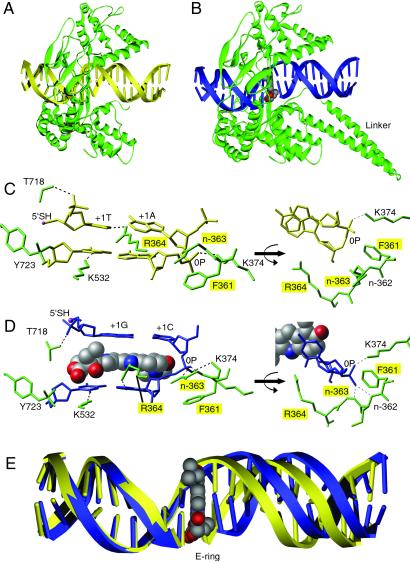
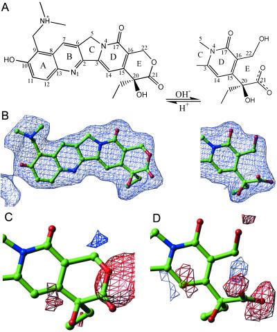
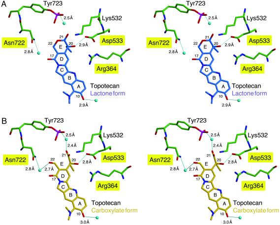
We report the x-ray crystal structure of human topoisomerase I covalently joined to double-stranded DNA and bound to the clinically approved anticancer agent Topotecan. Topotecan mimics a DNA base pair and binds at the site of DNA cleavage by intercalating between the upstream (-1) and downstream (+1) base pairs. Intercalation displaces the downstream DNA, thus preventing religation of the cleaved strand. By specifically binding to the enzyme-substrate complex, Topotecan acts as an uncompetitive inhibitor. The structure can explain several of the known structure-activity relationships of the camptothecin family of anticancer drugs and suggests that there are at least two classes of mutations that can produce a drug-resistant enzyme. The first class includes changes to residues that contribute to direct interactions with the drug, whereas a second class would alter interactions with the DNA and thereby destabilize the drug-binding site.


Figures

Fig 1.
Fig 1.
Structure of topo I–DNA complex without (A) and with (B) bound Topotecan. The binary complex is diagrammed with protein (green) and DNA (yellow). The ternary complex is diagrammed with protein (green), DNA (blue), and Topotecan (CPK gray as carbon, red as oxygen, blue as nitrogen). The 2.1-Å drug-bound structure represents the most complete topo I structure reported to date, providing visible electron density from Gln-201 to the COOH-terminal Phe-765. The linker domain (Pro-636 to Lys-712) could not be visualized in the binary complex (22) but was visible in the ternary complex. Previously reported crystal structures of human topo I include the inactive Y723F version of topo70 and topo58/6.3 (a reconstituted linkerless enzyme) in noncovalent complex with DNA and topo58/6.3 in covalent complex with DNA (13, 17). Unlike topo70, the reconstituted enzyme has altered kinetics and is not sensitive to CPT in a plasmid relaxation assay (17). (C) Molecular diagram showing the nondrug-bound topo I–DNA complex. The +1 and −1 bases of the duplex DNA (yellow, stick) are shown making four contacts to the surrounding protein (green, stick) to stabilize the protein–DNA complex. (D) Topotecan (CPK) intercalates between the +1 and −1 bases of the duplex DNA (blue, stick). Six protein (green, stick) contacts stabilize the open form of the DNA. A contact with the main chain nitrogen of Arg-362 is not labeled. Topo70 residues whose mutation leads to drug resistance are highlighted with yellow boxes. The 5′-SH of the +1 G is highlighted in pink. The covalent phosphotyrosine attachment to DNA is shown between Tyr-723 (green, stick) and the −1 T (blue, stick) of the cleaved strand. The mobile phosphodiester of the intact DNA strand is labeled 0P. (E) Comparison of the 22-mer duplex oligonucleotides of the drug-bound (blue) and nondrug-bound (yellow) complexes reveals that Topotecan (CPK) binds to the enzyme–substrate complex by intercalating in the DNA and shifting the downstream bases by ≈3.6 Å, equivalent to the rise of one base pair.
Fig 2.
Fig 2.
Topotecan electron density. (A) Topotecan with reversible hydrolysis of the E-ring from the closed lactone form to the open carboxylate form is diagrammed. (B) A 3.0 σ |Fo|–|Fc| omit map of electron density for Topotecan is illustrated. (C) A 3.0 σ |Fo|–|Fc| electron density map calculated with the lactone form of Topotecan (100% closed E-ring) is diagrammed. Negative electron density (red) is seen in the vicinity of the lactone oxygen, and positive (blue) electron density peaks are located nearby. (D) A 3.0 σ |Fo|–|Fc| electron density calculated with the carboxylate form of Topotecan (100% open E-ring) is diagrammed. Negative electron density (red) surrounds the terminal hydroxyl and carboxylic acid moieties, whereas a positive (blue) electron density peak is in the location of what would be the lactone oxygen in the closed E-ring conformation.
Fig 3.
Fig 3.
Stereoview of Topotecan binding. The Topotecan interactions with protein side chains for the lactone (A, blue) and carboxylate (B, gold) forms of the drug are depicted. Hydrogen bonds (predicted by contact distance and geometry of the refined atomic positions) are shown as solid blue lines, and the calculated interatomic distances are indicated. Labels for residues that result in resistance to CPT when mutated are highlighted in yellow. The oxygen atoms of water molecules are depicted as light blue spheres. Protein side chains are green, and noncarbon atoms are colored red for oxygen, blue for nitrogen, and magenta for phosphorus. The DNA and protein side chains that do not interact with Topotecan have been omitted from the figure.

References

    1. Wang J. C. (1996) Annu. Rev. Biochem. 65 635-692. - PubMed
    1. Champoux J. J. (2001) Annu. Rev. Biochem. 70 369-413. - PubMed
    1. Pommier Y. (1998) Biochimie 80 255-270. - PubMed
    1. Wall M. E., Wani, M. C., Cook, C. E., Palmer, K. H., McPhail, A. T. & Sim, G. A. (1966) J. Am. Chem. Soc. 88 3888-3890.
    1. Hsiang Y. H., Hertzberg, R., Hecht, S. & Liu, L. F. (1985) J. Biol. Chem. 260 14873-14878. - PubMed

Publication types

MeSH terms

LinkOut - more resources