Comparative Study
. 2002 Nov 26;99(24):15333-8.
doi: 10.1073/pnas.242501899. Epub 2002 Nov 8.

Tissue factor deficiency causes cardiac fibrosis and left ventricular dysfunction

Affiliations
Comparative Study

Tissue factor deficiency causes cardiac fibrosis and left ventricular dysfunction

R Pawlinski et al. Proc Natl Acad Sci U S A. .

Abstract

Exposure of blood to tissue factor (TF) activates the extrinsic (TF:FVIIa) and intrinsic (FVIIIa:FIXa) pathways of coagulation. In this study, we found that mice expressing low levels of human TF ( approximately 1% of wild-type levels) in an mTF(-/-) background had significantly shorter lifespans than wild-type mice, in part, because of spontaneous fatal hemorrhages. All low-TF mice exhibited a selective heart defect that consisted of hemosiderin deposition and fibrosis. Direct intracardiac measurement demonstrated a 30% reduction (P < 0.001) in left ventricular function in 8-month-old low-TF mice compared with age-matched wild-type mice. Mice expressing low levels of murine FVII ( approximately 1% of wild-type levels) exhibited a similar pattern of hemosiderin deposition and fibrosis in their hearts. In contrast, FIX(-/-) mice, a model of hemophilia B, had normal hearts. Cardiac fibrosis in low-TF and low-FVII mice appears to be caused by hemorrhage from cardiac vessels due to impaired hemostasis. We propose that TF expression by cardiac myocytes provides a secondary hemostatic barrier to protect the heart from hemorrhage.


Figures

Fig 1.
Fig 1.
Survival of low-TF mice on mixed and C57BL/6J backgrounds. Kaplan–Meier plots showing survival profiles of 77 low-TF mice on a mixed background (Mixed) and 61 low-TF mice on a C57BL/6J background (C57).
Fig 2.
Fig 2.
Fibrosis in hearts of low-TF mice. (A) The degree of fibrosis was scored (0–5) in heart sections stained with Masson's Trichrome. Fibrosis was scored in hearts from 35 mice on a mixed background and 31 hearts on a C57BL/6J background. (B) Cross-sections of hearts stained with Masson's Trichrome demonstrate subepicardial and myocardial fibrosis in the hearts of low-TF mice (8 and 23 months) and no fibrosis in control mice (8 months). Normal myocardium stains red-brown and fibrotic tissue stains blue. Original magnification of the panels from the cross-sections was ×100. Hearts shown are from low-TF mice on a mixed genetic background. LV, left ventricle.
Fig 3.
Fig 3.
LV function in the hearts of low-TF mice. LV function was performed on 8-month-old low-TF mice (n = 6) on a C57BL/6J background and age-matched C57BL/6J mice (n = 5). (Left) LV function was measured at different heart rates. (Center and Right) LV function and LV pressure at normal heart rates (480–510 beats per min).
Fig 4.
Fig 4.
Hemosiderin deposits in the hearts of low-TF mice. Serial sections of a low-TF mouse on a mixed background (8 months of age) were stained with H&E (A), Prussian Blue (B), and Masson's Trichrome (C). Hemosiderin appears as a golden-brown deposit with H&E and stains blue with Prussian Blue. (Original magnification ×400.)
Fig 5.
Fig 5.
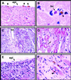
Histological analysis of the hearts of low-TF mice. Heart sections were stained with H&E. (Original magnifications: A, ×100; B, ×1,000; C, ×250; DF, ×400.) Hearts of low-TF mice (line 47) at 3 months (A), 5 months (C), 8 months (B and D), 11 months (F), and 14 months (E) of age are shown. Hemosiderin (brown) is observed subepicardially (A) and perivascularly (B). (C and D) Interstitial hemorrhages and necrosis of a cardiac myocyte (nuclear pyknosis and loss of cross striations; arrow; D). (E) Leukocyte infiltration and cardiac myocyte necrosis. (F) Fibrosis (light pink) and associated hemosiderin deposition with some remaining cardiac myocytes (dark pink). Hearts shown are from low-TF mice on a mixed background. Blood vessels (bv), epicardium (epi), and hemosiderin (arrowheads) are indicated.
Fig 6.
Fig 6.
Analysis of hearts of mice with deficiencies in the extrinsic pathway of coagulation. Heart sections were stained with Prussian Blue (AC) or Masson's Trichrome (D). Shown are heart sections from an mTF−/−/hTF+(line 47) mouse (12 weeks of age; A), an mTF−/−/hTF+(line 31) mouse (8 weeks of age; B), and an FVIItTA-FVII/tTA-FVII mouse (9 weeks of age; C and D).
Fig 7.
Fig 7.
Model showing how TF contributes to primary and secondary hemostatic barriers in cardiac and skeletal muscle. Skeletal muscle contains only a primary TF hemostatic barrier surrounding blood vessels (dark pink), whereas cardiac muscle contains both primary and secondary (light pink) TF hemostatic barriers. Endothelial cell layer is shown in green and blood is shown in red.

References

    1. Drake T. A., Morrissey, J. H. & Edgington, T. S. (1989) Am. J. Pathol. 134 1087-1097. - PMC - PubMed
    1. Edgington T. S., Mackman, N., Brand, K. & Ruf, W. (1991) Thromb. Haemostasis 66 67-79. - PubMed
    1. Davie E. W., Fujikawa, K. & Kisiel, W. (1991) Biochemistry 30 10363-10370. - PubMed
    1. Hogan K. A., Weiler, H. & Lord, S. T. (2002) Thromb. Haemostasis 87 563-574. - PubMed
    1. Christie P. D., Edelberg, J. M., Picard, M. H., Foulkes, A. S., Mamuya, W., Weiler-Guettler, H., Rubin, R. H., Gilbert, P. & Rosenberg, R. D. (1999) J. Clin. Invest. 104 533-539. - PMC - PubMed

Publication types

MeSH terms

LinkOut - more resources