. 2002 Dec;35(6):321-32.
doi: 10.1046/j.1365-2184.2002.00240.x.

Nitric oxide inhibits irreversibly P815 cell proliferation: involvement of potassium channels

Affiliations

Nitric oxide inhibits irreversibly P815 cell proliferation: involvement of potassium channels

R S A Costa et al. Cell Prolif. 2002 Dec.

Abstract

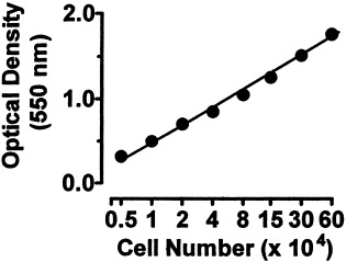
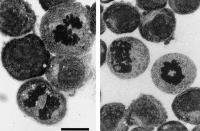
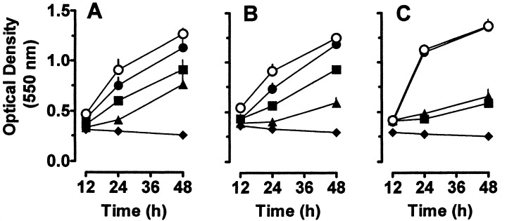
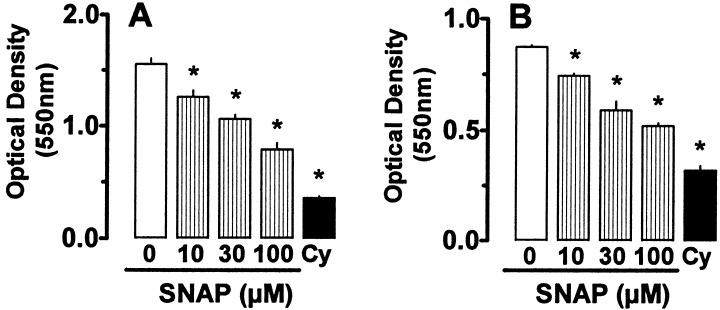
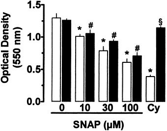
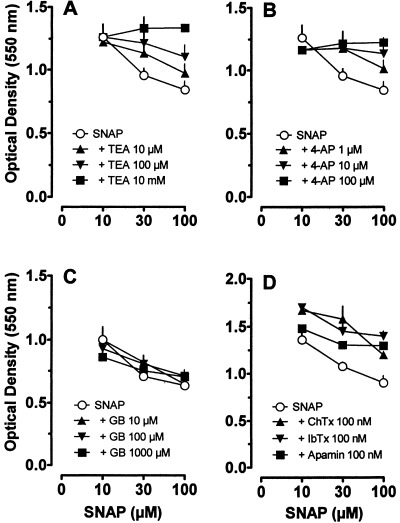
Nitric oxide (NO) has been shown to inhibit both normal and cancer cell proliferation. Potassium channels are involved in cell proliferation and, as NO activates these channels, we investigated the effect of NO on the proliferation of murine mastocytoma cell lines and the putative involvement of potassium channels. NO (in the form of NO donors) caused dose-dependent inhibition of cell proliferation in the P815 cell line inducing growth arrest in the mitosis phase. Incubation with NO donor for 4 or 24 h had a similar inhibitory effect on cell proliferation, indicating that this effect is irreversible. The inhibitory effect of NO was completely prevented by the blockade of voltage- and calcium-dependent potassium channels, but not by blockade of ATP-dependent channels. NO inhibition of cell proliferation was unaffected by guanylate cyclase and by cytoskeleton disruptors. Therefore, NO inhibits cell proliferation irreversibly via a potassium channel-dependent but guanylate cyclase-independent pathway in murine mastocytoma cells.


Figures

Figure 1
Figure 1
Validation of the crystal violet method in comparison with counting in Neubauer chambers. Known numbers of P815 viable cells (determined in Neubauer chambers) were plated in triplicate wells of a 96‐well plate in 200 µl medium. Each group was then fixed and stained with crystal violet solution, washed with distilled water, homogenized in SDS 1% (w/v) and centrifuged. The optical density was read at 550 nm in a plate reader. Each point represents the mean ± SEM of triplicates. When no error bar is visible, it was covered by the symbol. The x axis is in log scale and the correlation coefficient was 0.886.
Figure 2
Figure 2
SNAP arrests P815 cells in mitosis. Cells were incubated with SNAP (100 µ m ) for 24 h and the mitotic figures were counted in slides stained with May‐Grünwald‐Giemsa as described in Materials and Methods section. Photographs were taken under oil immersion. Bar represents 20 µm, total magnification × 1000.
Figure 3
Figure 3
Concentration‐ and time‐dependent effect of NO donors on P815 cell proliferation. Cells (5 × 10 4 cells/well; 200 µl medium) were seeded and incubated with NO donors or medium for the indicated time periods. Then, cultures were fixed and stained with crystal violet solution, washed, homogenized and the optical density was read at 550 nm, as detailed in Materials and Methods section. In all panels, open circles represent cells incubated with medium alone and the closed diamonds represent cells incubated with cycloheximide (18 µ m ). (a) SNAP at 10 (closed circles), 30 (squares) or 100 µ m (triangles). (b) GSNO at 100 (closed circles), 300 (squares) or 1000 µ m (triangles). (c) SNP at 1 (closed circles), 10 (squares) or 100 µ m (triangles). Points represent mean ± SEM of triplicates. When no error bar is visible, it was covered by the symbol. Similar results were obtained in at least two identical experiments. All curves in closed symbols were statistically different from the control curve, as determined by one‐way analysis of variance ( anova ) followed by Bonferroni's posthoc t ‐test.
Figure 4
Figure 4
Effect of SNAP on the proliferation of P815 and MCP‐5 mastocytoma cell lines. P815 cells (a) and MCP‐5 (b) (5 × 10 4 cells/well; 200 µl medium) were seeded and incubated with SNAP at the indicated concentrations, cycloheximide (Cy; 18 µ m ) or medium alone for 24 h. Cells were then fixed and stained with crystal violet solution, washed, homogenized and the optical density was read at 550 nm. Similar results were obtained in at least two identical experiments. Bars are mean ± SEM of triplicate wells. The symbol * means significant differences ( P  < 0.05) from each respective control group, using one‐way analysis of variance ( anova ) followed by Bonferroni's posthoc t ‐test.
Figure 5
Figure 5
Irreversibility of SNAP‐induced inhibition of proliferation in P815 cells. Cells (5 × 10 4 cells/well; 200 µl medium) were seeded and incubated with SNAP, cycloheximide (Cy; 18 µ m ) or medium for 4 h (closed bars) or for 24 h (open bars). Cultures were then fixed, stained with crystal violet solution, washed, homogenized and the optical density was read at 550 nm, as detailed in Materials and Methods section. Each bar represents mean ± SEM of triplicates. Similar results were obtained in at least two identical experiments. The symbols * and # mean significant differences ( P  < 0.05) from each respective control group and the symbol § means significant difference compared to cycloheximide incubated for 4 h, using one‐way analysis of variance ( anova ) followed by Bonferroni's posthoc t ‐test.
Figure 6
Figure 6
Effect of potassium channel blockers on SNAP‐induced inhibition of P815 cell proliferation. Cells (5 × 10 4 cells/well; 200 µl medium) were seeded and incubated with the compounds or medium for 24 h. Cultures were then fixed and stained with crystal violet solution, washed, homogenized and the optical density was read at 550 nm, as detailed in Materials and Methods section. In all panels, open circles represent the effect of SNAP alone at the indicated concentrations. (a) SNAP plus tetraethylammonium (TEA) at 10 µ m (triangles), 100 µ m (inverted triangles) and 10 m m (squares); (b) SNAP plus 4‐aminopyridine (4‐AP) at 1 µ m (triangles), 10 µ m (inverted triangles) and 100 µ m (squares); (c) SNAP plus glibenclamide (GB) at 10 µ m (triangles), 100 µ m (inverted triangles) and 1000 µ m (squares); (d) SNAP plus charybdotoxin (ChTx) at 100 n m (triangles); SNAP plus iberiotoxin (IbTx) at 100 n m (inverted triangles) and SNAP plus apamin at 100 n m (squares). Symbols represent mean ± SEM of triplicates. When no error bar is visible, it was covered by the symbol. Similar results were obtained in at least two identical experiments. All curves in closed symbols are statistically different ( P  < 0.05) from SNAP control curve (open symbols), except those depicted in (c) (SNAP plus glibenclamide), as determined by one‐way analysis of variance ( anova ) followed by Bonferroni's posthoc t ‐test.

Similar articles

References

    1. Albina JE, Reichner JS (1998) Role of nitric oxide in mediation of macrophage cytotoxicity and apoptosis. Cancer Metastasis Rev. 17, 39. - PubMed
    1. Arnelle DR, Stamler JS (1995) NO+, NO· and NO− donation by S‐nitrosothiols: implications for regulation of physiological functions by S‐nitrosylation and acceleration of disulfide formation. Arch. Biochem. Biophys. 318, 279. - PubMed
    1. Beckman JS, Koppenol WH (1996) Nitric oxide, superoxide, and peroxynitrite: the good, the bad, and the ugly. Am. J. Physiol. 271, C1424. - PubMed
    1. Bolotina VM, Najibi S, Palacino JJ, Pagano PJ, Cohen RA (1994) Nitric oxide directly activates calcium‐dependent potassium channels in vascular smooth muscle. Nature 362, 850. - PubMed
    1. Broillet MC (1999) S‐Nitrosylation of proteins. Cell Mol. Life Sci. 55, 1036. - PMC - PubMed

Publication types

MeSH terms

LinkOut - more resources