. 2002 Nov 12;2(1):3.
doi: 10.1186/1471-2342-2-3.

CT features in abdominal tuberculosis: 20 years experience

Affiliations

CT features in abdominal tuberculosis: 20 years experience

Tariq Sinan et al. BMC Med Imaging. .

Abstract

BACKGROUND: Abdominal tuberculosis (TB) is endemic in the developing world and is reemerging in the West. Since computed tomography (CT) has the ability to demonstrate changes in the peritonium, mesentry, lymphnodes, bowel and solid organs and is being increasingly used for primary evaluation of abdominal conditions, it is important to be familiar with the CT features of the disease. METHODS: CT findings were retrospectively analysed in 49 patients with proved abdominal TB. Patients with genitourinary TB and with AIDS/HIV were not included in the study. RESULTS: Peritoneal involvement was the most common feature (77.5%) with ascites (wet peritonitis) seen in more than half the cases (55.2%). The rest showed peritoneal, mesenteric or omental thickening or mass formation but no ascites (dry peritonitis). Other findings included lymphadenopathy (46.9% mainly of diffuse nature, bowel wall thickening (38%) and solid organ involvement (20.4%). CONCLUSIONS: CT reliably demonstrates the entire range of findings which need interpretation in the light of clinical and laboratory data.


Figures

Figure 1
Figure 1
Wet peritonitis: CT scan of the pelvis showing large volume free ascites. Note omental mass (arrows).
Figure 2
Figure 2
Wet peritonitis: CT scan mid-abdomen showing small volume loculated ascites (thick arrow) Note mesenteric strands and mesenteric nodes (thin arrow). Peritoneal thickening is seen in the right side.
Figure 3
Figure 3
Dry peritonitis: CT scan showing diffuse mesenteric strands, mesenteric nodes and omental thickening.
Figure 4
Figure 4
Circumferencial thickening of the caecum and narrowing of the terminal ilium. Also note mesenteric strands and omental thickening.
Figure 5
Figure 5
Necrotic lymph nodes with lucent center seen in the (a) portal/peri pancreatic region (arrow) and (b) para-aortic area.
Figure 6
Figure 6
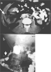
(a) CT scan showing circumferential thickening of the ascending colon (short arrow). Note right parietal abscess with free air (long arrow). (b) This was due to a fistula (arrow) of the ascending colon confirmed by barium enema.
Figure 7
Figure 7
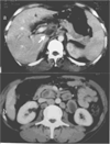
CT of the upper abdomen showing large necrotic inflammatory mass in the lesser sac and involving the pancreas. This was due to perforating gastric TB ulcer.
Figure 8
Figure 8
CT of the upper abdomen showing multiple focal hypodense lesions in the liver.
Figure 9
Figure 9
CT of the upper abdomen focal lesions in the spleen. Note associated para aortic nodes (arrows).

Similar articles

Cited by

References

    1. Dye C, Scheele S, Dolin P, Pathania V, Raviglione MC. Consensus statement. Global burden of tuberculosis: estimated incidence, prevalence, and mortality by country. WHO Global Surveillance and Monitoring Project. JAMA. 1999;282:677–686. doi: 10.1001/jama.282.7.677. - DOI - PubMed
    1. Suri S, Gupta S, Suri R. Computed tomography in abdominal tuberculosis. B J Radiol. 1999;72:92–8. - PubMed
    1. Jadvar H, Mindelzun RE, Olcott EW, Levitt DB. Still the great mimicker: Abdominal tuberculosis. AJR Am J Roentgenol. 1997;168:1455–60. - PubMed
    1. Akhan O, Pringot J. Imaging of abdominal tuberculosis. Eur Radiol. 2002;12:312–23. doi: 10.1007/s003300100994. - DOI - PubMed
    1. Batra A, Gulati MS, Sarma D, Paul SB. Sonographic appearances in abdominal tuberculosis. J of Clin Ultrasound. 2000;28:233–245. doi: 10.1002/(SICI)1097-0096(200006)28:5<233::AID-JCU5>3.0.CO;2-C. - DOI - PubMed

LinkOut - more resources