

Reinterpretation of cross-sectional images in patients with head and neck cancer in the setting of a multidisciplinary cancer center
- PMID: 12427610
- PMCID: PMC8185819
Reinterpretation of cross-sectional images in patients with head and neck cancer in the setting of a multidisciplinary cancer center
Abstract
Background and purpose: Patients referred to tertiary care centers frequently arrive with images obtained at outside institutions; these images require reinterpretation. We assessed the clinical value of reinterpreting cross-sectional imaging studies of patients with head and neck cancer, in the setting of a multidisciplinary cancer center.
Methods: Outside CT and MR images of 136 patients with known or presumed head and neck cancer were reinterpreted by a neuroradiologist. Clinical history and findings on physical examination were available. Reinterpretation was performed before review of outside reports, which were subsequently compared with those generated at the cancer center. Changes in interpretation were noted, and their effects on TNM staging, patient care, and prognosis were assessed. Reliability and statistical significance of rates of change in diagnosis were analyzed with 95% confidence intervals (CIs) and the sign test, respectively. Verification of change in diagnosis was confirmed by pathologic analysis (75%), characteristic radiologic findings (18%), or clinical and imaging follow-up (7%).
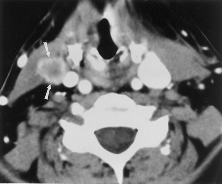
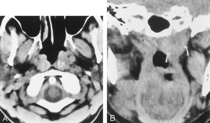
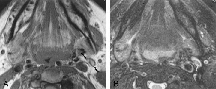
Results: Change in interpretation occurred in 56 patients (41%) (95% CI: 33-49%, P <.001). Forty-six patients (34%) had a change in T, N, and/or M staging (26-42%, P <.001). Change in T stage occurred in 27 cases (20%) (13-27%, P <.001) (upstaged in 22, downstaged in five), and a change in N stage in 26 cases (19%) (12-26%, P <.001) (upstaged in 20, downstaged in six). Two patients (1.5%) had missed systemic metastases. Three patients with an initial diagnosis of cancer were found to be cancer-free, and six patients had a diagnosis of new second primary cancers that were missed at original interpretation. One patient had a missed middle cerebral artery aneurysm. Changes in image interpretation altered treatment in 55 (98%) of 56 patients and affected prognosis in 53 patients (95%) (P <.001).
Conclusion: Reinterpretation of cross-sectional images in the setting of a multidisciplinary cancer center has a significant effect on staging, management, and prognosis in patients with head and neck cancer.
Figures
Comment in
-
Reinterpretation of head and neck scans: massive can of worms or call to action?AJNR Am J Neuroradiol. 2002 Nov-Dec;23(10):1617-8. AJNR Am J Neuroradiol. 2002. PMID: 12427606 Free PMC article. No abstract available.
References
-
- Kazerooni NL, Kazerooni EA, Quint LE, Orringer MB. Added value of thoracic radiology specialist interpretation of CT scans for lung cancer consultations in a thoracic surgery clinic (abstr). Radiology 1998;209(P):171
-
- Gollub MJ, Panicek DM, Bach AM, Penalver BS, Castellino RA. Clinical importance of reinterpretation of body CT scans obtained elsewhere in patients referred for care at a tertiary cancer center. Radiology 1999;210:109–112 - PubMed
-
- Kalbhen CL, Yetter EM, Love L, Moncada R, Lawson TL, Albain KA. Outside film reviews of thoracic imaging studies in oncology patients (abstr). AJR Am J Roentgenol 1998;170(suppl):77
-
- Hricak H, Kalbhen CL, Scheidler JE, Schwartz LH, Yu KK, Adams D. Value of expert interpretation in abdominal oncologic imaging: a multicenter study (abstr). Radiology 1997;205(P):225
-
- Bechtold RE, Chen MYM, Ott DJ, et al. Interpretation of abdominal CT: analysis of errors and their causes. J Comput Assist Tomogr 1997;21:681–685 - PubMed
Publication types
MeSH terms
LinkOut - more resources
Full Text Sources
Medical