

Fragile X premutation carriers: characteristic MR imaging findings of adult male patients with progressive cerebellar and cognitive dysfunction
- PMID: 12427636
- PMCID: PMC8185834
Fragile X premutation carriers: characteristic MR imaging findings of adult male patients with progressive cerebellar and cognitive dysfunction
Abstract
Background and purpose: Our purpose was to characterize the findings of MR imaging of the brain of adult male fragile X premutation carriers with a recently identified disorder characterized by ataxia, tremor, rigidity, and cognitive dysfunction.
Methods: MR imaging studies of the brain of 17 male patients were characterized for signal intensity and for size of ventricles, cerebral and cerebellar sulci, and brain stem. Comparison was made with age- and sex-matched control participants. Southern blot and/or polymerase chain reaction methods were used to analyze CGG trinucleotide repeats in the fragile X mental retardation 1 gene.
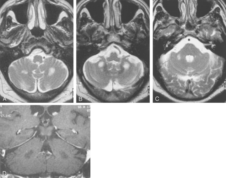
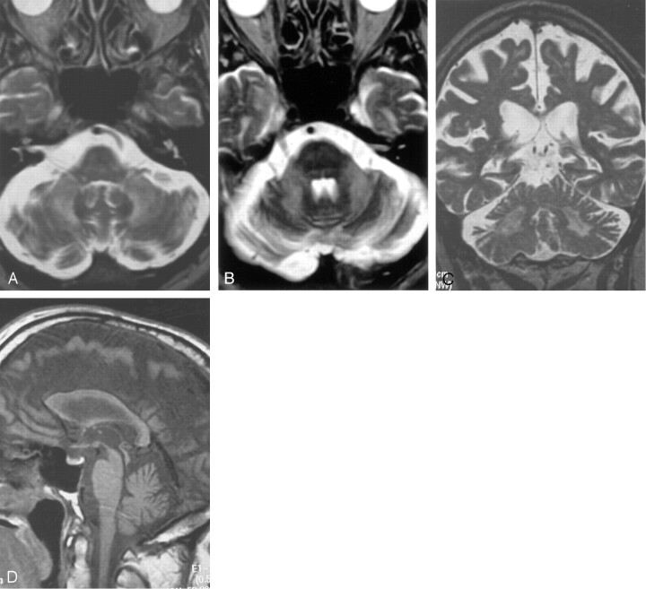
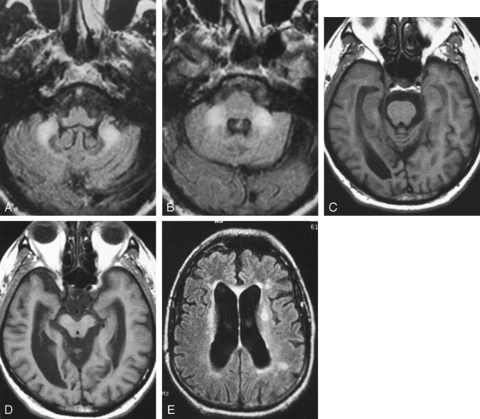
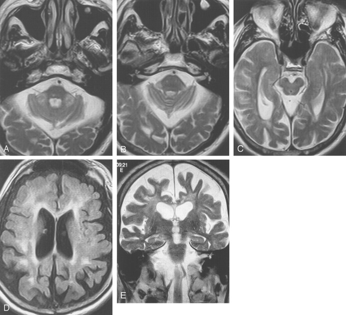
Results: Fifteen of 17 patients showed symmetrically decreased T1 and increased T2 signal intensity in cerebellar white matter lateral, superior, and inferior to the dentate nuclei. Fourteen of 17 had similar signal intensity alterations in the middle cerebellar peduncles. Cerebellar cortical atrophy was present in 16 of 17 and cerebral atrophy in 17 of 17. Evan's Index as a measure of ventricular size averaged 0.35 (range, 0.25-0.46), with that for age-matched control participants averaging 0.28 (range, 0.24-0.31) (P <.005). The mean third ventricle width was 11 mm (for control participants, 6 mm; P <.01). Corpus callosum was thinned in 14 of 16 participants. Middle cerebellar peduncles were atrophic when compared with those of control participants (P <.005). Pontine transverse dimension was 25 mm (for control participants, 31 mm; P <.005), and rostral-caudal length averaged 26 mm (for control participants, 29 mm; P <.005). CGG repeats clustered in the low to mid premutation range (86 +/- 10 CGG repeats) in the 17 patients.
Conclusion: MR imaging findings in symptomatic male fragile X premutation carriers are characteristic of this disorder. Recognition of these alterations may support a specific diagnosis and may have implications for the potential occurrence of fragile X syndrome in the children of reproductive age female relatives.
Figures
References
-
- Rousseau F, Morel M-L, Rouillard P, Khandjian EW, Morgan K. Surprisingly low prevalence of FMR1 premutation among males from the general population. American Society of Human Genetics Annual Meeting, October 28–November 1,1997. , Baltimore.
-
- Hagerman RJ, Hagerman PJ. Fragile X syndrome: a model of gene-brain-behavior relationships. Mol Genet Metab 2001;74:89–97 - PubMed
-
- Dorn MB, Mazzocco MM, Hagerman RJ. Behavioral and psychiatric disorders in adult male carriers of fragile X. J Am Acad Child Adolesc Psychiatry 1994;256–264 - PubMed
-
- Franke P, Leboyer M, Gansicke M, et al. Genotype-phenotype relationship in female carriers of the premutation and full mutation of FMR-1. Psych Res 1998;80:113–127 - PubMed
Publication types
MeSH terms
Substances
Grants and funding
LinkOut - more resources
Full Text Sources
Other Literature Sources
Medical