. 2002 Nov 15;22(22):9679-86.
doi: 10.1523/JNEUROSCI.22-22-09679.2002.

Two distinct signaling pathways upregulate NMDA receptor responses via two distinct metabotropic glutamate receptor subtypes

Affiliations

Two distinct signaling pathways upregulate NMDA receptor responses via two distinct metabotropic glutamate receptor subtypes

Pascal Benquet et al. J Neurosci. .

Abstract

Molecular processes regulating the gain of NMDA receptors modulate diverse physiological and pathological responses in the CNS. Group I metabotropic glutamate receptors (mGluRs), which neighbor NMDA receptors and which can be coactivated by synaptically released glutamate, couple to several different second messenger pathways, each of which could target NMDA receptors. In CA3 pyramidal cells we show that the activation of mGluR1 potentiates NMDA current via a G-protein-independent mechanism involving Src kinase activation. In contrast, mGluR5-mediated enhancement of NMDA current requires G-protein activation, triggering a signaling cascade including protein kinase C and Src. These results indicate that one neurotransmitter, glutamate, can activate two distinct and independent signaling systems to target the same effector. These two pathways are likely to contribute significantly to the highly differentiated control of NMDA receptor function.


Figures

Fig. 1.
Fig. 1.
Activation of group I mGluRs potentiates currents mediated by NMDA receptors in CA3 pyramidal cells. A, NMDA currents induced by the pressure application of NMDA (100 μm for 200 msec) every 40 sec (blackarrows) are potentiated by the bath application of the specific group I mGluR agonist DHPG (10 μm for 2 min). The effect is transient and reversible. B, Single NMDA current traces from the recording in A shown at an expanded time scale before (1) during (2), and after (3) the washout of DHPG. C, Average time course of the DHPG-induced potentiation (n = 23).D, Average current–voltage relationship of NMDA responses obtained with a ramp protocol before (filled circles) and after (open circles) DHPG application (n = 5; mean ± SEM of current is shown every 2 mV). Inset shows subtraction of controlI–V plot from I–V plot after the application of DHPG.
Fig. 2.
Fig. 2.
Src is required for mGluR-mediated potentiation of NMDA current. A, Genistein, a broad-spectrum blocker of tyrosine kinase, inhibits DHPG-induced potentiation of NMDA current.A1, Single traces from the same neuron show that the increase in NMDA current amplitude induced by DHPG (10 μm) is prevented in the presence of genistein (30 μm for 15 min). Genistein alone does not alter NMDA current. A2, Averaged results from five cells comparing the effect on NMDA current of 10 μm DHPG alone (open circles) and in the presence of genistein (filledcircles).A3, Potentiation of NMDA current by 10 μmDHPG is inhibited by genistein (30 μm;n = 5) but not by its inactive analog daidzein (30 μm;n = 5); *p < 0.05. B, PP1, a specific inhibitor of Src kinase, inhibits the DHPG-induced potentiation of NMDA current.B1, Time course of action on peak NMDA current of DHPG (10 μm for 2 min) before and after a 15 min application of 25 μm PP1 in a representative cell. PP1 alone does not alter NMDA current. B2, Single NMDA current traces from this cell. B3, Averaged results from five cells comparing the effect on NMDA current of DHPG before (open circles) and after (filled circles) PP1 incubation. B4, Pooled data comparing responses to DHPG alone and DHPG in the presence of PP1 (n = 5); *p < 0.05. Dotted lines indicate baseline or control responses.
Fig. 3.
Fig. 3.
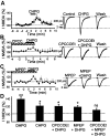
Activation of either mGluR5 or mGluR1 potentiates NMDA current. A, Average time course of NMDA current potentiation induced by the mGluR5-specific agonist CHPG (500 μm for 2 min; n = 8). Representative traces from one cell are shown (right).B, Alternatively, mGluR5 was stimulated selectively by the application of DHPG (10 μm for 2 min) in the presence of a saturating concentration of the mGluR1 antagonist CPCCOEt (50 μm for 10 min; n = 6). Average time course (left) and representative traces (right) are shown. C, mGluR1 was stimulated selectively by the application of DHPG (10 μmfor 2 min) in the presence of a saturating concentration of the mGluR5 antagonist MPEP (10 μm for 10 min). Average time course (left) and representative traces (right) are shown. D, Pooled data indicate that, although the selective activation of either mGluR5 (CHPG, n = 8; CPCCOEt plus DHPG, n = 4) or mGluR1 (MPEP plus DHPG, n = 5) significantly potentiates NMDA current, greater potentiation is observed with the coactivation of mGluR1 and mGluR5 (DHPG, n = 23). No significant potentiation is detected in the presence of both mGluR1 and mGluR5 antagonists (CPCCOEt plus MPEP plus DHPG, n = 5); *p < 0.05 and **p < 0.01.
Fig. 4.
Fig. 4.
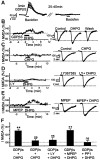
G-protein-independent potentiation of NMDA current is mediated by mGluR1 but not by mGluR5. A, Within 3 min of establishing the whole-cell configuration with a patch pipette containing 1 mm GDPβS, the application of baclofen (20 μm for 1 min) induces an outward K+current. Having allowed 20–40 min for GDPβS to diffuse into the cell, reapplication of baclofen no longer produces a response, indicating a blockade of the G-protein function (n= 6). B, After the baclofen response is blocked completely, DHPG (10 μm for 2 min) still potentiates NMDA current (n = 6). Representative traces from one cell are shown on the right. C, Average time course of NMDA current in six GDPβS-treated cells exposed to the mGluR5-specific agonist CHPG (500 μm for 2 min), indicating a lack of potentiation. Representative traces from one cell are shown on the right. D, Average time course of NMDA current in five GDPβS-treated cells in which mGluR5 activation is obtained by applying DHPG (10 μm for 2 min) in the presence of a saturating concentration of the mGluR1 antagonist LY367385 (50 μm for 10 min), again showing that NMDA current is not potentiated. Representative traces from one cell are shown on the right. E, Average time course of NMDA current in five GDPβS-treated cells in which mGluR1 is activated selectively by applying DHPG (10 μm for 2 min) in the presence of a saturating concentration of the mGluR5 antagonist MPEP (10 μm for 10 min), showing marked potentiation. Representative traces from one cell are shown on theright. F, Pooled data for GDPβS-treated cells showing that the activation of mGluR1 plus mGluR5 with DHPG (n = 6) significantly potentiates NMDA current. Selective activation of mGluR5 with CHPG (n = 6) or LY367385 plus DHPG (n = 5) does not potentiate NMDA current, whereas the selective activation of mGluR1 (MPEP plus DHPG,n = 5) potentiates NMDA current to a similar degree, as does the coactivation of mGluR1 plus mGluR5 with DHPG. DHPG-induced potentiation is blocked completely by the Src inhibitor PP1 (25 μm for 20 min; n = 5); **p < 0.01.
Fig. 5.
Fig. 5.
Blocking PKC activation prevents NMDA current potentiation by mGluR5 but not by mGluR1. For all experiments with the PKC inhibitor, GDPβS was not present and BAPTA (10 mm) was used in the pipette solution to minimize the Ca2+-dependent inhibition of NMDA current.A, Left, Average time course of NMDA current potentiation induced by DHPG (10 μm for 2 min) either with EGTA (open circles; 10 mm;n = 23) or with BAPTA (filled circles; 10 mm;n = 6) in the recording pipette. A, Right, Data using the same protocol but with the mGluR5-specific agonist CHPG (500 μm for 2 min) either with EGTA (10 mm;n = 8) or with BAPTA (10 mm;n = 7) in the recording pipette. B, Inhibition of PKC with the specific inhibitor GF109203X (2 μm for 20 min) prevents NMDA current potentiation in response to the mGluR5-specific agonist CHPG (500 μm for 2 min; n = 6). Representative traces from one cell are shown on the right. C, Similarly, inhibition of PKC prevents NMDA current potentiation in response to mGluR5 activation by the application of DHPG (10 μm for 2 min; n = 4) in the presence of a saturating concentration of the mGluR1 antagonist CPCCOEt (50 μm for 10 min). Representative traces from one cell are shown on theright. D, In contrast, the selective activation of mGluR1 by the application of DHPG (10 μmfor 2 min) in the presence of a saturating concentration of the mGluR5 antagonist MPEP (10 μm for 10 min) still potentiates NMDA current under conditions in which PKC is blocked (n= 5). Representative traces from one cell are shown on theright. E, Average time course of NMDA current potentiation induced by DHPG (10 μm for 2 min;n = 5) in the presence of the PKC inhibitor. Representative traces from one cell are shown on theright. F, Pooled data showing that, after the inhibition of PKC, the selective activation of mGluR5 (CHPG,n = 6; or LY367385 plus DHPG, n = 4) does not potentiate NMDA current, whereas the selective activation of mGluR1 (MPEP plus DHPG, n = 5) induces a potentiation comparable with that seen with the coactivation of mGluR1 plus mGluR5. Note that NMDA current potentiation induced by the coactivation of mGluR1 plus mGluR5 after PKC inhibition is reduced but still significant (n = 5); *p < 0.05.

References

    1. Adler EM, Augustine GJ, Duffy SN, Charlton MP. Alien intracellular calcium chelators attenuate neurotransmitter release at the squid giant synapse. J Neurosci. 1991;11:1496–1507. - PMC - PubMed
    1. Alagarsamy S, Sorensen SD, Conn PJ. Coordinate regulation of metabotropic glutamate receptors. Curr Opin Neurobiol. 2001;11:357–362. - PubMed
    1. Ali DW, Salter MW. NMDA receptor regulation by Src kinase signaling in excitatory synaptic transmission and plasticity. Curr Opin Neurobiol. 2001;11:336–342. - PubMed
    1. Aniksztejn L, Bregestovski P, Ben-Ari Y. Selective activation of quisqualate metabotropic receptor potentiates NMDA but not AMPA responses. Eur J Pharmacol. 1991;205:327–328. - PubMed
    1. Anwyl R. Metabotropic glutamate receptors: electrophysiological properties and role in plasticity. Brain Res Brain Res Rev. 1999;29:83–120. - PubMed

Publication types

MeSH terms

LinkOut - more resources