. 2002 Nov 15;22(22):9868-76.
doi: 10.1523/JNEUROSCI.22-22-09868.2002.

Localization of phosphorylated cAMP response element-binding protein in immature neurons of adult hippocampus

Affiliations

Localization of phosphorylated cAMP response element-binding protein in immature neurons of adult hippocampus

Shin Nakagawa et al. J Neurosci. .

Abstract

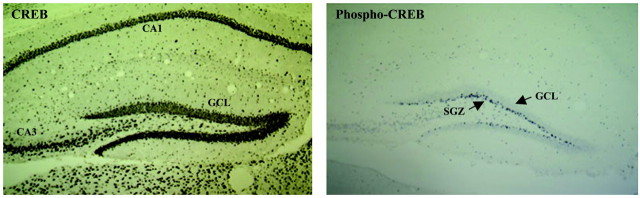
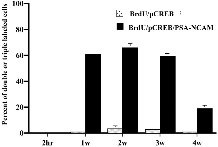
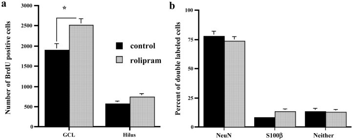
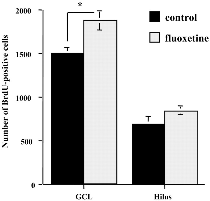
Neurogenesis continues to occur in the adult hippocampus, although many of the newborn cells degenerate 1-2 weeks after birth. The number and survival of newborn cells are regulated by a variety of environmental stimuli, but very little is known about the intracellular signal transduction pathways that control adult neurogenesis. In the present study, we examine the expression of the phosphorylated cAMP response element-binding protein (pCREB) in immature neurons in adult hippocampus and the role of the cAMP cascade in the survival of new neurons. The results demonstrate that virtually all immature neurons, identified by triple immunohistochemistry for bromodeoxyuridine (BrdU) and polysialic acid-neural cell adhesion molecule (PSA-NCAM), are also positive for pCREB. In addition, upregulation of cAMP (via pharmacological inhibition of cAMP breakdown or by antidepressant treatment) increases the survival of BrdU-positive cells. A possible role for pCREB in the regulation of PSA-NCAM, a marker of immature neurons involved in neuronal remodeling and neurite outgrowth, is supported by cell culture studies demonstrating that the cAMP-CREB pathway regulates the expression of a rate-limiting enzyme responsible for the synthesis of PSA-NCAM. These findings indicate that the cAMP-CREB pathway regulates the survival, and possibly the differentiation and function, of newborn neurons.


Figures

Fig. 1.
Fig. 1.
The distribution of CREB (a) and phosphorylated CREB (b) in the adult mouse dentate gyrus. CREB is phosphorylated constitutively in the cells near the subgranular zone, whereas CREB is expressed in almost all cells of hippocampus, including the CA1 and CA3 pyramidal cell layers and the dentate gyrus granule cell layer.
Fig. 2.
Fig. 2.
Colocalization of CREB or pCREB and PSA-NCAM in the developing BrdU-positive cells. a–c,Top, Representative confocal micrographs depicting double immunolabeling for BrdU (green) and CREB (red) 2 hr after BrdU injection. Thepanels below depict triple immunostaining for BrdU (blue), PSA-NCAM (green), and pCREB (red) at different time points after BrdU injection: 2 hr (d–g), 2 weeks (h–k), or 4 weeks (l–o). Yellowarrows, Localization of BrdU-positive cells. Note that BrdU-positive cells migrate from the subgranular zone into granule cell layer at the later time points and transiently express pCREB and PSA-NCAM at the 2 week, but not the 2 hr or 4 week, time point. Scale bars: c, o, 20 μm.
Fig. 3.
Fig. 3.
Time course for colocalization of pCREB and/or PSA-NCAM in BrdU-positive cells in the adult hippocampus. Triple immunolabeling was conducted as described in Materials and Methods and Figure 2. The number of cells expressing pCREB and BrdU or pCREB, BrdU, and PSA-NCAM was determined by identifying 50 BrdU-positive cells for each animal (n = 5) at each time point. Thex-axis indicates the time after BrdU injection. The results are presented as percentage of double- or triple-labeled cells and are the mean ± SEM (n = 250 at each time point).
Fig. 4.
Fig. 4.
Colocaliation of TUC-4 and pCREB in the developing BrdU-positive cells. Representative confocal micrographs are shown for triple labeling of BrdU (blue), TUC-4 (green; TOAD-64), and pCREB (red) at different time points after BrdU injections: 2 hr (a–d) or 2 weeks (e,f). Yellow arrows, Localization of BrdU-positive cells. Two weeks after injection, BrdU-positive cells are colabeled with TUC-4 and pCREB. Scale bar, 20 μm.
Fig. 5.
Fig. 5.
Influence of rolipram administration on the number (a) and phenotype (b) of surviving BrdU-positive cells in the adult hippocampus. Mice were injected with BrdU on 4 consecutive days to label newborn cells. One week after the beginning of BrdU injections (3 d after the last injection), animals were administered rolipram once daily for 3 weeks as described in Materials and Methods. a, The number of BrdU-positive cells throughout the entire hippocampus was determined using a modified unbiased stereological procedure. The results are expressed as the number of BrdU-positive cells in the granule cell layer or hilus of the bilateral dentate gyrus (mean ± SEM). *p < 0.01 compared with vehicle-treated controls (Student's t test). b, The percentage of surviving BrdU-positive cells stained with NeuN, a marker of mature neurons, or S100β, a glial marker, was also determined. There was no significant difference in the phenotype of the BrdU-positive cells between control and chronic rolipram-treated mice.
Fig. 6.
Fig. 6.
Influence of chronic fluoxetine administration on the number of surviving BrdU-labeled cells in hippocampus. Animals were administered BrdU and were then treated with either vehicle or fluoxetine for 4 weeks as indicated. Analysis of the number of BrdU-positive cells was conducted 4 weeks after the first BrdU administration. The number of BrdU-positive cells throughout the entire hippocampus was determined using a modified stereological procedure as described in Materials and Methods. The results are expressed as the mean ± SEM number of BrdU-positive cells in the granule cell layer or hilus of the entire dentate gyrus. *p < 0.01 compared with vehicle-treated controls (Student'st test).
Fig. 7.
Fig. 7.
Influence of the cAMP–CREB cascade on STX promoter activity. Deletion mutants of the STX promoter, shown ina, were transfected into PC12 cells, which were then incubated with either vehicle (Control) or forskolin (10 μm), and levels of luciferase reporter activity were determined (b). The influence of mCREB on STX promoter activity was determined (c). The 0.8 kb construct, which contains the putative CRE, and a construct containing mCREB were cotransfected into PC12 cells. The cells were incubated with vehicle (Control) or forskolin, and levels of luciferase activity were determined. The results (b,c) are expressed as a percentage of the activity of the control plasmid (lacking insert) and are the mean of three separate experiments, each performed in duplicate.

References

    1. Barad M, Bourtchouladze R, Winder DG, Golan H, Kandel E. Rolipram, a type IV-specific phosphodiesterase inhibitor, facilitates the establishment of long-lasting long-term potentiation and improves memory. Proc Natl Acad Sci USA. 1998;95:15020–15025. - PMC - PubMed
    1. Becker C, Artola A, Gerardy-Schahn R, Becker T, Welzl H, Schachner M. The polysialic acid modification of the neural cell adhesion molecule is involved in spatial learning and hippocampal long-term potentiation. J Neurosci Res. 1996;45:143–152. - PubMed
    1. Bender R, Lauterborn JC, Gall CM, Cariaga W, Baram TZ. Enhanced CREB phosphorylation in immature dentate gyrus granule cells precedes neurotrophin expression and indicates a specific role of CREB in granule cell differentiation. Eur J Neurosci. 2001;13:679–686. - PMC - PubMed
    1. Biebl M, Cooper CM, Winkler J, Kuhn HG. Analysis of neurogenesis and programmed cell death reveals a self-renewing capacity in the adult rat brain. Neurosci Lett. 2000;291:17–20. - PubMed
    1. Bonfani L, Olive S, Poulain DA, Theodosis DT. Mapping of the distribution of polysialylated neural cell adhesion molecule throughout the central nervous system of the adult rat: an immunohistochemical study. J Neurosci. 1992;19:10228–10236. - PubMed

Publication types

MeSH terms