. 2002 Nov 15;22(22):9885-94.
doi: 10.1523/JNEUROSCI.22-22-09885.2002.

Thalamocortical bursts trigger recurrent activity in neocortical networks: layer 4 as a frequency-dependent gate

Affiliations

Thalamocortical bursts trigger recurrent activity in neocortical networks: layer 4 as a frequency-dependent gate

Michael Beierlein et al. J Neurosci. .

Abstract

Sensory information reaches the cortex via thalamocortical (TC) synapses in layer 4. Thalamic relay neurons that mediate information flow to cortex operate in two distinct modes, tonic and burst firing. Burst firing has been implicated in enhancing reliability of information flow between individual neurons. However, little is known about how local networks of neocortical neurons respond to different temporal patterns of TC activity. We studied cortical activity patterns evoked by stimulating TC afferents at different frequencies, using a combination of electrophysiology and calcium imaging in TC slices that allowed for the reconstruction of spatiotemporal activity with single-cell resolution. Stimulation of TC axons at low frequencies triggered action potentials in only a small number of layer 4 neurons. In contrast, brief high-frequency stimulus trains triggered widespread recurrent activity in populations of neurons in layer 4 and then spread into adjacent layers 2/3 and 5. Recurrent activity had a clear threshold, typically lasted 300 msec, and could be evoked repetitively at frequencies up to 0.5 Hz. Moreover, the spatial extent of recurrent activity was controlled by the TC pattern of activity. Recurrent activity triggered within the highly interconnected networks of layer 4 might act to selectively amplify and redistribute transient high-frequency TC inputs, filter out low-frequency inputs, and temporarily preserve a record of past sensory activity.


Figures

Fig. 1.
Fig. 1.
Imaging TC-evoked activity in TC slices.A, Schematic of TC slice, with location of approximate recording site in layer 4. B, Neocortical layer 4 viewed with 20× objective. Slice was loaded with fura-2 AM. Outlined area is shown at higher magnification in the right panel. C, D, Single TC stimulus evokes response in single neuron. C, Normalized fluorescence change for neuron (solid line) shown inB and D and entire field of view (dotted line). Vertical arrow in this and all subsequent figures indicates time of TC stimulation.D, Fluorescence change is limited to single neuron. Shown is one frame (320 msec duration) of ΔF/F0 movie, corresponding to time after the stimulus. Activated neuron is marked by anarrow in both B andD.
Fig. 2.
Fig. 2.
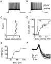
TC EPSPs trigger spikes in layer 4 excitatory neurons. All data except B recorded from neuron shown in Figure 1. A, B, Intrinsic firing pattern of RS neurons in layer 4. Depolarizing current steps (0.1 nA) evoked spike firing with either fast (A) or slow (B) degree of spike adaptation. C, TC responses in RS cell identified via Ca2+ signal. Stimulus amplitude was adjusted to evoke threshold responses, with an approximately equal number of successes and failures. Multiple trials evoked at 0.1 Hz are superimposed. D, Spike latency jitter in an RS cell. Shown are four consecutive trials evoked at 0.1 Hz. E, Dependence of spike latency on membrane potential in an RS cell. Holding potential was adjusted by small depolarizing or hyperpolarizing currents (<20 pA). AP, Action potential. F, Small number of TC axons mediates spiking. Incrementing stimulus intensity results in stepwise increase in EPSP amplitude. Cell was hyperpolarized by ∼5 mV to prevent spiking.
Fig. 3.
Fig. 3.
Rapid feedforward inhibition is mediated by FS interneurons. A, B, Intrinsic firing in two types of interneurons evoked by depolarizing current steps (0.2 pA). A, FS cell, with no spike frequency adaptation.B, RSNP cell with moderate spike frequency adaptation. C, Precise spike generation in FS neurons triggered by TC EPSPs. Raster plot indicates time of action potential firing in consecutive trials (0.1 Hz) evoked by single TC stimuli applied at time 0. D, Distribution of spike latencies for FS and RS cells, evoked by single TC EPSPs. Shown are the median values for each cell. E, Small number of TC axons evokes spikes in FS neuron. Incrementing stimulus intensity results in stepwise increase in EPSP amplitude. Cell was hyperpolarized by ∼5 mV to prevent spiking. F, Feedforward inhibition curtails TC EPSPs. Recording from an RS cell, held at −55 mV. Multiple trials are overlaid.
Fig. 4.
Fig. 4.
High-frequency bursts trigger recurrent activity.A, Network activity in layer 4 evoked by short train of TC stimuli (5 stimuli, 40 Hz). Leftmost frame shows imaged area in layer 4, viewed by 20× objective. Three consecutive frames of ΔF/F0 movie (320 msec duration per frame), with frame 1 preceding the stimulus burst and frames 2 and 3following the stimulus burst. B, Imaging lateral spread of network activity at low magnification. Slice viewed with 4× objective. Network activity evoked by three stimuli, 40 Hz. Three consecutive frames of ΔF/F0movie (200 msec duration per frame), with frame 1preceding the stimulus burst and frames 2 and3 following the stimulus burst. Initiation of activity in layer 4 (frame 2) is followed by lateral spread in layers 2/3 and 5 and restricted spread in layer 4 (frame 3). C, Network activity is mediated by intracortical polysynaptic activity. Recordings show synaptic responses from two RS neurons in the same barrel, evoked by four stimuli at 10 Hz (left) and 40 Hz (right). Cell 2 did not receive TC EPSPs but was recruited by polysynaptic activity. D, Facilitating monosynaptic EPSP in an FS cell, evoked by thalamic stimulation (8 stimuli, 40 Hz). Note the absence of recurrent activity.
Fig. 5.
Fig. 5.
Initiation of recurrent activity in layer 4.A, Leftmost frame shows imaged area in layer 4. Activity evoked by 40 Hz stimulus train of four (top row) or five pulses (bottom row). Three consecutive frames of ΔF/F0movie (320 msec duration) are shown for both stimulus trains. Notice that the number and spatial extent of initially activated neurons (frame 1) are similar, whereas only longer trains evoke recurrent activity (frames 2 and3, bottom row). Graph plots normalized fluorescence change in imaged area during entire movie, evoked by 40 Hz trains of three, four, or five pulses. Arrow marks onset of stimulus train. B, Stimulus dependence of recurrent activity. Increasing number of pulses beyond the threshold of recurrent activity recruits additional neurons. Shown are three frames (320 msec duration) of three separate ΔF/F0 movies corresponding to the time frame after the respective stimulus train at 40 Hz (left, 3 pulses; middle, 4 pulses;right, 5 pulses). Initiation site of population activity is to the left of imaged area.
Fig. 6.
Fig. 6.
Recurrent activity is blocked by reducing NMDA-mediated EPSPs. A, Activity evoked by four stimuli at 40 Hz, before (top row) and 5 min after (bottom row) application of 2 μm APV . For each case, three consecutive frames of ΔF/F0 movie (320 msec duration per frame) are shown, with frame 1 preceding the stimulus burst and frames 2 and 3following the stimulus burst. B, APV completely blocks recurrent activity and slightly attenuates TC EPSPs. Shown are responses in an FS cell, evoked every 2 min by TC stimulus train (8 stimuli, 20 Hz) after the application of 2 μm APV. Notice the increasing delay of recurrent activity before failure. Spikes are truncated.
Fig. 7.
Fig. 7.
Near-linear recruitment of layer 4 neurons by TC stimulus trains. Activity was evoked in the presence of 2 μm APV. Shown are three frames (320 msec duration) of three separate ΔF/F0 movies corresponding to the time frame after the respective stimulus train at 40 Hz for four, five, and six pulses. Notice the gradual increase of activity, without significant spread into layers 2/3 and 5. B, Graph plots peak normalized fluorescence change in imaged area, under control conditions and after APV application as shown in A, evoked by stimulus trains of three to six pulses at 40 Hz.
Fig. 8.
Fig. 8.
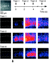
Population activity can be evoked repetitively. Recurrent activity was evoked by six consecutive stimulus trains (3 stimuli, 40 Hz) at 0.5 Hz. Shown are cortical activity patterns after the first, second, and sixth stimulus train. Frame duration, 200 msec. Notice the persistence of activity in layer 4 and the larger response variability in layers 2/3.

References

    1. Agmon A, Connors BW. Thalamocortical responses of mouse somatosensory (barrel) cortex in vitro. Neuroscience. 1991;41:365–379. - PubMed
    1. Agmon A, Yang LT, O'Dowd DK, Jones EG. Organized growth of thalamocortical axons from the deep tier of terminations into layer IV of developing mouse barrel cortex. J Neurosci. 1993;13:5365–5382. - PMC - PubMed
    1. Agmon A, Hollrigel G, O'Dowd DK. Functional GABAergic synaptic connection in neonatal mouse barrel cortex. J Neurosci. 1996;16:4684–4695. - PMC - PubMed
    1. Ahmed B, Anderson JC, Douglas RJ, Martin KA, Nelson JC. Polyneuronal innervation of spiny stellate neurons in cat visual cortex. J Comp Neurol. 1994;341:39–49. - PubMed
    1. Chen C, Regehr WG. Developmental remodeling of the retinogeniculate synapse. Neuron. 2000;28:955–966. - PubMed

Publication types

MeSH terms

LinkOut - more resources