. 2002 Nov 15;22(22):9912-21.
doi: 10.1523/JNEUROSCI.22-22-09912.2002.

Mechanisms of amygdala modulation of hippocampal plasticity

Affiliations

Mechanisms of amygdala modulation of hippocampal plasticity

Irit Akirav et al. J Neurosci. .

Abstract

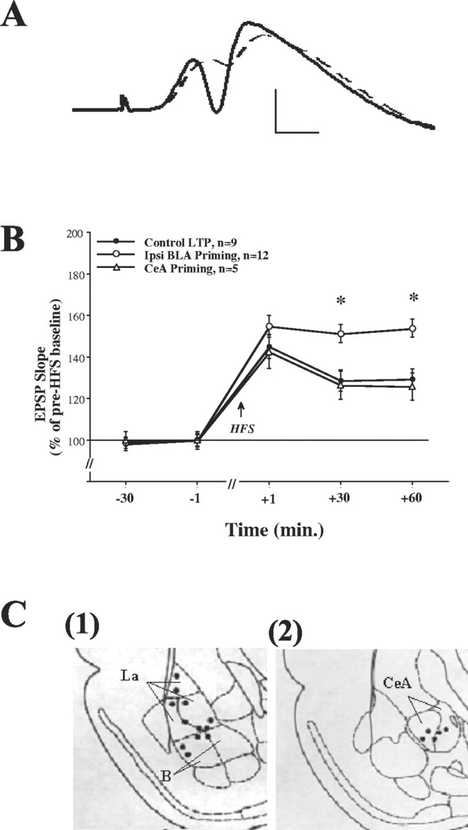
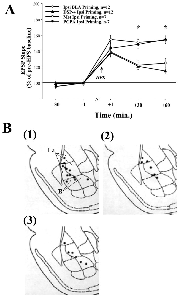
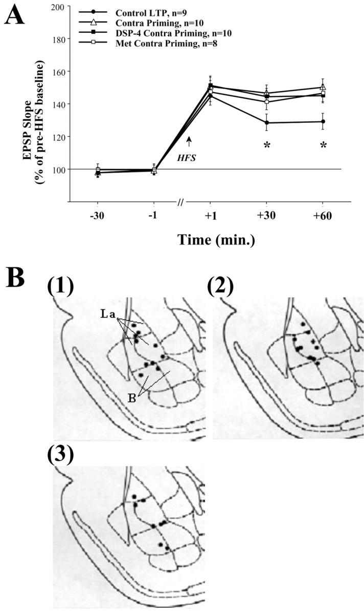
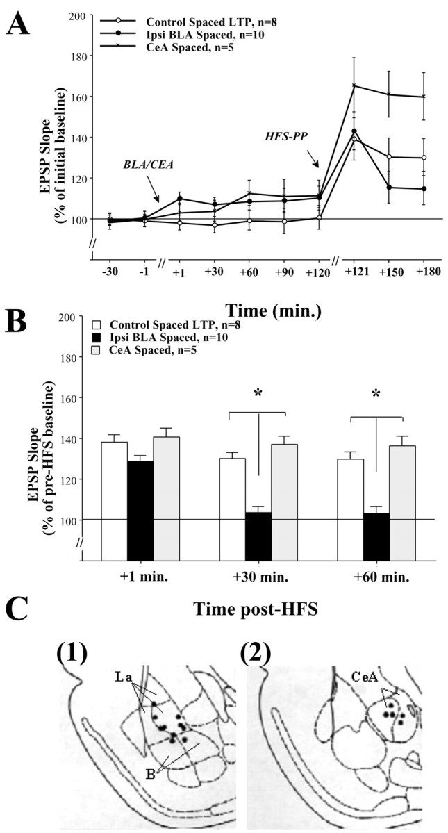
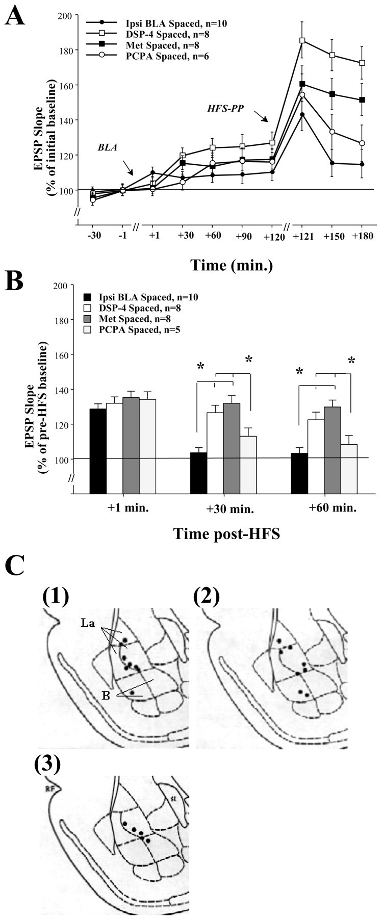
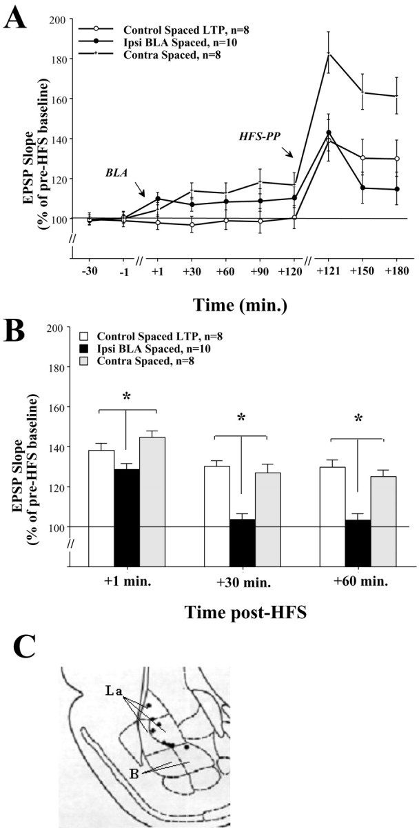
Basolateral amygdala (BLA) activation by emotional arousal modulates memory-related processes in the hippocampus. We have shown (Akirav and Richter-Levin, 1999b) that activating the BLA before perforant path (PP) tetanization has a biphasic effect on hippocampal plasticity; priming the BLA immediately before PP tetanization results in the enhancement of dentate gyrus (DG) long-term potentiation (LTP) (an "emotional tag"), whereas stimulation in a spaced interval results in the suppression of DG-LTP. Here, we aimed to elucidate the mechanisms underlying BLA modulation of DG-LTP and specifically to examine whether the stress hormones norepinephrine (NE) and corticosterone (CORT) are main mediators of the BLA biphasic effects. We found that the BLA affects hippocampal plasticity in a complex manner; BLA priming enhanced DG-LTP, and both NE and CORT mediated this effect. Furthermore, we found that ipsilateral BLA spaced activation (2 hr before PP tetanization) suppressed DG-LTP and that this suppressive effect was also mediated by NE and CORT. Priming the contralateral BLA enhanced DG-LTP similarly to the ipsilateral enhancement, but neither NE nor CORT mediated this effect. The spaced activation of the contralateral BLA did not suppress DG-LTP. Taken together, these results suggest that differential mechanisms underlie the ipsilateral and contralateral BLA effects on hippocampal plasticity. Hence, the BLA modulates hippocampal memory processes, presumably via the mediation of the stress hormones NE and CORT, to establish a diverse memory of the experience. Possibly, at the onset of an emotional event the stress hormones permissively mediate plasticity. However, their prolonged presence in the system may suppress the cognitive response to stress.


Figures

Fig. 1.
Fig. 1.
A, Representative evoked potentials recorded from the DG before (dashed line) and after HFS to the PP. Calibration: vertical, 5 mV; horizontal, 1 msec.B, Ipsilateral BLA priming enhances DG-LTP, whereas CeA priming does not. The increase in EPSP slope (LTP) was measured as a percentage of baseline value immediately before HFS to the PP. The levels of DG-LTP in the Control LTP group (n = 9) after HFS were significantly different from 100% at all times post-HFS (see Results). Ipsilateral priming stimulation of the BLA (Ipsi BLA Priming, n=12) induced 30 sec before HFS was applied to the PP significantly increased DG-LTP levels compared with the Control LTP group at +30 min (*p < 0.001) and at +60 min post-HFS (*p < 0.0001), confirming our previous reports of BLA priming enhancing effect on DG-LTP (Akirav and Richter-Levin, 1999a,b). Priming the CeA (CeA Priming, n=5) did not enhance DG-LTP levels compared with the Control LTP group, and the CeA Priming group was significantly different from the Ipsi BLA Priming group at both +30 min (*p < 0.01) and +60 min post-HFS (*p < 0.0001). This suggests that the CeA does not modulate DG plasticity.C, Schematic drawings of BLA and CeA electrode placements. Shown is a coronal view at positions 3.14 and 2.56 mm posterior to bregma for the BLA and the CeA, respectively. Solid black circles indicate the locations: (1) Ipsi BLA Priming group and (2) CeA Priming group (B, basal amygdala; La, lateral amygdala; together they form the BLA).
Fig. 2.
Fig. 2.
A, Ipsilateral BLA priming is mediated by NE and CORT but not by 5-HT. Priming the BLA in NE-depleted (DSP-4 Ipsi Priming, n=12) or CORT-depleted (Met Ipsi Priming, n=7) rats did not enhance DG-LTP as seen in the Ipsi BLA Priming group (+30 min: DSP-4 Ipsi Priming, *p < 0.0001; Met Ipsi Priming, p < 0.01; +60 min: DSP-4 Ipsi Priming, *p < 0.0001; Met Ipsi Priming, p < 0.001). This suggests that both NE and CORT may mediate the BLA priming enhancing effect on hippocampal LTP. In contrast, priming the BLA in 5-HT-depleted rats (PCPA Ipsi Priming, n=7) enhanced DG-LTP as in the Ipsi BLA Priming group, and this group was significantly different from the other depleted groups at both +30 min (DSP-4 Ipsi Priming, *p < 0.01; Met Ipsi Priming, p < 0.01) and +60 min post-HFS (DSP-4 Ipsi Priming, *p < 0.0001;Met Ipsi Priming, p < 0.01). This suggests that the BLA priming enhancing effect on DG-LTP is not dependent on serotonergic activation. B, Schematic drawings of BLA electrode placements. Solid black circles indicate the locations: (1) DSP-4 Ipsi Priming group, (2) Met Ipsi Priming group, and (3) PCPA Ipsi Priming group.
Fig. 3.
Fig. 3.
A, Contralateral priming enhances DG-LTP but is not NE or CORT dependent. Priming the contralateral BLA (Contra Priming, n=10) significantly enhanced DG-LTP levels compared with the control LTP group at both +30 min (*p < 0.01) and +60 min post-HFS (*p < 0.001). In addition, there was no significant difference between the Contra Priming group and the Ipsi BLA Priming group shown in Figure 1A. This suggests that priming the contralateral BLA enhances DG-LTP in a way similar to the enhancement seen by ipsilateral priming. Priming the contralateral BLA of rats depleted of NE (DSP-4 Contra Priming, n=10) or CORT (Met Contra Priming, n=8) also resulted in enhanced DG-LTP levels. They were significantly different from the control LTP group at both +30 min (Control LTP, *p < 0.01; DSP-4 Contra Priming,p < 0.05), and +60 min post-HFS (Control LTP, *p < 0.01; DSP-4 Contra Priming, p < 0.01) but were not different from the Contra Priming group. This suggests that, although contralateral BLA priming enhances DG-LTP, this effect is NE and CORT independent. B, Schematic drawings of BLA electrode placements. Solid black circles indicate the locations: (1) Contra Priming group, (2) DSP-4 Contra Priming group, and (3) Met Contra Priming group.
Fig. 4.
Fig. 4.
A, DG-EPSP slope after BLA and CeA stimulation. Shown is the DG-EPSP slope (in response to PP stimulation) for the duration of the experiment. B, Ipsilateral BLA spaced activation suppresses DG-LTP, whereas CeA spaced activation does not. The levels of DG-LTP in the Control Spaced LTP group (n = 8) after HFS to the PP were significantly different from 100% at all the times post-HFS to the PP (+1, +30, and + 60 min) (see Results). Spaced stimulation of the BLA (Ipsi BLA Spaced, n=10) significantly reduced DG-LTP levels compared with the Control Spaced LTP group at +30 min (*p < 0.0001) and +60 min post-HFS (*p < 0.0001). This confirms our previous reports showing BLA spaced activation suppressing DG-LTP levels (Akirav and Richter-Levin, 1999b). Spaced activation of the CeA (CeA Spaced, n=5) did not reduce DG-LTP levels compared with the Control Spaced LTP group and was significantly different from the Ipsi BLA Spaced group at both +30 min (*p < 0.0001) and +60 post-HFS (*p < 0.0001). This suggests that under these conditions CeA spaced activation does not modulate DG-LTP. Note that, although BLA and CeA spaced activation induced a shift in baseline EPSP (A), the levels of LTP in the CeA group were similar to that in the control group. This indicates that the lack of potentiation in the BLA spaced group was not caused by saturation of plasticity as a result of the shift in baseline EPSP before HFS.C, Schematic drawings of BLA and CeA electrode placements. Solid black circles indicate the locations: (1) Ipsi BLA Spaced group and (2) CeA Spaced group.
Fig. 5.
Fig. 5.
A, DG-EPSP slope after BLA stimulation in NE-, CORT-, and 5-HT-depleted animals. Shown is the DG-EPSP slope (in response to PP stimulation) for the duration of the experiment. B, Ipsilateral BLA spaced activation is mediated by NE and CORT but not by 5-HT. BLA spaced activation in NE-depleted (DSP-4 Spaced, n=8) or CORT-depleted (Met Spaced, n=8) rats did not reduce DG-LTP levels as seen in the Ipsi BLA Spaced rats at both +30 min (DSP-4 Spaced, *p < 0.0001; Met Spaced, *p < 0.0001) and +60 min post-HFS (DSP-4 Spaced, *p < 0.0001;Met Spaced, p < 0.01). Moreover, the NE-and CORT-depleted groups were not significantly different from the Control Spaced LTP group shown in Figure 4B. This suggests that both NE and CORT may mediate the BLA spaced suppressing effect on DG-LTP. BLA spaced activation in 5-HT-depleted rats (PCPA Spaced, n=6) significantly reduced DG-LTP compared with the NE- and CORT-depleted rats at both +30 min (DSP-4 Spaced, *p < 0.05;Met Spaced, p < 0.01) and +60 min post-HFS (DSP-4 Spaced, *p < 0.05;Met Spaced, p < 0.01). Moreover, there was no significant difference between the PCPA Spaced group and the Ipsi BLA Spaced group at any time point. This suggests that the BLA spaced suppressing effect on DG-LTP is not dependent on serotonergic activation. C, Schematic drawings of BLA electrode placements. Solid black circles indicate the locations: (1) DSP-4 Spaced group, (2) Met Spaced group, and (3) PCPA Spaced group.
Fig. 6.
Fig. 6.
A, DG-EPSP slope after contralateral BLA stimulation. Shown is the DG-EPSP slope (in response to PP stimulation) for the duration of the experiment.B, Contralateral spaced activation does not suppress DG-LTP. Contralateral BLA spaced activation (n = 8) did not suppress DG-LTP as seen in the Ipsi BLA Spaced group at +1 min (*p < 0.01), +30 min (*p < 0.001), and +60 min post-HFS (*p < 0.001), and the Contra BLA Spaced group was not significantly different from the Control Spaced LTP group. Together with the results from Figure3A, this further supports the possibility that the ipsilateral and contralateral BLA effects on DG-LTP are mediated via different mechanisms. C, Schematic drawings of BLA electrode placements. Solid black circles indicate the locations of the Contra BLA Spaced group.

References

    1. Abe K. Modulation of hippocampal long-term potentiation by the amygdala: a synaptic mechanism linking emotion and memory. Jpn J Pharmacol. 2001;86:18–22. - PubMed
    1. Akirav I, Richter-Levin G. Priming stimulation in the basolateral amygdala modulates synaptic plasticity in the rat dentate gyrus. Neurosci Lett. 1999a;30:83–86. - PubMed
    1. Akirav I, Richter-Levin G. Biphasic modulation of hippocampal plasticity by behavioral stress and basolateral amygdala stimulation in the rat. J Neurosci. 1999b;1:10530–10535. - PMC - PubMed
    1. Amorapanth P, LeDoux JE, Nader K. Different lateral amygdala outputs mediate reactions and actions elicited by a fear-arousing stimulus. Nat Neurosci. 2000;3:74–79. - PubMed
    1. Bhatnagar S, Costall B, Smythe JW. Hippocampal cholinergic blockade enhances hypothalamic-pituitary-adrenal responses to stress. Brain Res. 1997;22:244–248. - PubMed

Publication types

MeSH terms