. 2002 Nov;130(3):1241-53.
doi: 10.1104/pp.011742.

Characterization of AtCDC48. Evidence for multiple membrane fusion mechanisms at the plane of cell division in plants

Affiliations

Characterization of AtCDC48. Evidence for multiple membrane fusion mechanisms at the plane of cell division in plants

David M Rancour et al. Plant Physiol. 2002 Nov.

Abstract

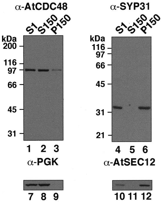
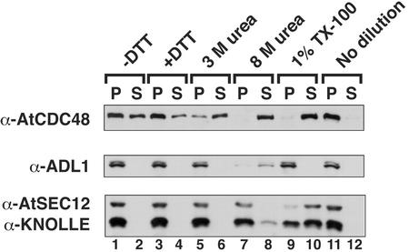
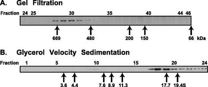
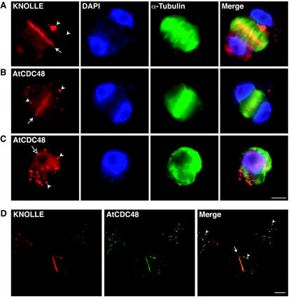
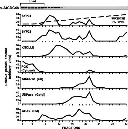
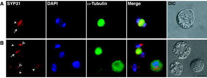
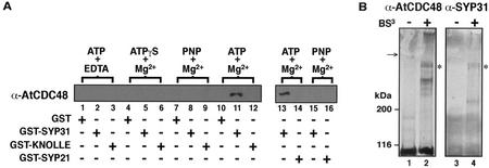
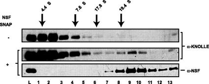
The components of the cellular machinery that accomplish the various complex and dynamic membrane fusion events that occur at the division plane during plant cytokinesis, including assembly of the cell plate, are not fully understood. The most well-characterized component, KNOLLE, a cell plate-specific soluble N-ethylmaleimide-sensitive fusion protein (NSF)-attachment protein receptor (SNARE), is a membrane fusion machine component required for plant cytokinesis. Here, we show the plant ortholog of Cdc48p/p97, AtCDC48, colocalizes at the division plane in dividing Arabidopsis cells with KNOLLE and another SNARE, the plant ortholog of syntaxin 5, SYP31. In contrast to KNOLLE, SYP31 resides in defined punctate membrane structures during interphase and is targeted during cytokinesis to the division plane. In vitro-binding studies demonstrate that AtCDC48 specifically interacts in an ATP-dependent manner with SYP31 but not with KNOLLE. In contrast, we show that KNOLLE assembles in vitro into a large approximately 20S complex in an Sec18p/NSF-dependent manner. These results suggest that there are at least two distinct membrane fusion pathways involving Cdc48p/p97 and Sec18p/NSF that operate at the division plane to mediate plant cytokinesis. Models for the role of AtCDC48 and SYP31 at the division plane will be discussed.


Figures

Figure 1
Figure 1
Specificity of AtCDC48 and SYP31 antibodies. Arabidopsis subcellular fractions (20 μg; S1, S150, and P150) were resolved by SDS-PAGE and analyzed by immunoblotting with AtCDC48 (lanes 1–3) and SYP31 (lanes 4–6) antibodies. Cytosolic PGK (lanes 7–9) and membrane-associated AtSEC12 (lanes 10–12) were used to confirm the identity and relative purity of each subcellular fraction.
Figure 2
Figure 2
AtCDC48 is peripherally associated with membranes. Microsomal membranes (P150) were diluted and incubated in the absence (lanes 1–2) or presence of 1 mm dithiothreitol (DTT; lanes 3–4), 3 m urea (lanes 5–6), 8 m urea (lanes 7–8), 1% (v/v) TX-100 (lanes 9–10), or no dilution was made (lanes 11–12). Samples were fractionated by differential centrifugation. Protein equivalents (20 μg) of soluble and pelletable material were analyzed by immunoblotting using antibodies directed against AtCDC48, Arabidopsis dynamin-like protein 1 (ADL1; peripheral), AtSEC12 (integral ER), and KNOLLE (integral SNARE). Protein recovery and loading was analyzed by PonceauS staining before immunoblot development.
Figure 3
Figure 3
Cytosolic AtCDC48 exists as a large heterogeneous protein complex. Arabidopsis cytosol (S150) was fractionated by Superose-6 HR 10/30 gel filtration chromatography (A) and glycerol velocity gradient sedimentation (B). Protein mobility standards (arrows) were run in parallel to the sample. Individual fractions were analyzed by immunoblotting and Coomassie staining to localize AtCDC48 or mass standards, respectively.
Figure 4
Figure 4
Localization of AtCDC48 in interphase and dividing Arabidopsis cells. Dividing Arabidopsis cells were analyzed by wide-field indirect immunofluorescence microscopy (A–C) and confocal microscopy (D). A through C, Cells were immunolabeled with anti-α-tubulin (green) and affinity-purified anti-KNOLLE (row A; red) or anti-AtCDC48 (rows B and C; red) antibodies and 4′,6-diamino-phenylindole (DAPI; blue). Electronically complied images (merged) were generated from the pseudocolored images. D, Colocalization (yellow) of KNOLLE (red) and AtCDC48 (green) in dividing (center) and nondividing (top left and right) cells were examined by indirect confocal immunofluorescence microscopy. White arrows indicate the location of the cell plate. White arrowheads indicate the position of subcellular membrane compartments containing KNOLLE and AtCDC48 (see text for discussion). The unfilled arrow (C) indicates nuclear localization of AtCDC48. Bar = 50 μm.
Figure 5
Figure 5
Membrane-bound AtCDC48 is primarily associated with a low-density membrane fraction. An Arabidopsis post-nuclear supernatant (S1) was fractionated by velocity centrifugation on a Suc gradient at 150,000g for 2 h at 4°C. The refractive index (expressed as Suc %, w/w) and content of various subcellular marker proteins of each gradient fraction was determined by enzyme activity assays (Golgi UDPase) or immunoblotting.
Figure 6
Figure 6
Localization of SYP31 in dividing Arabidopsis cells. Arabidopsis cells were double immunolabeled with anti-α-tubulin antibodies (green) and affinity-purified anti-SYP31 antibodies (red) and DAPI. Merged images were generated electronically from the three preceding pseudocolored images and are shown in the indicated panels (merged). Differential interphase contrast images of the cells analyzed are presented (DIC). White arrows indicate the location of the cell plate. White arrowheads indicate the position of large undefined subcellular structures. Unfilled arrowheads indicate the position of small cytoplasmic punctate structures. Bar = 50 μm.
Figure 7
Figure 7
AtCDC48 interacts with SYP31. A, AtCDC48 interacts with SYP31 in vitro. Bacterially expressed GST and GST fusion proteins containing the cytosolic domains of SYP31 (GST-SYP31), SYP21 (GST-SYP21), and KNOLLE (GST-KNOLLE) were incubated with Arabidopsis cytosol in the presence of adenosine nucleotides (1 mm) and either Mg2+ (1 mm) or EDTA (10 mm). Binding of AtCDC48 was assessed by immunoblotting. PNP is an abbreviation for 5′-adenylylimidodiphosphate. B, AtCDC48 interacts with SYP31 in vivo. An Arabidopsis post-nuclear supernatant (S1) was treated with the chemical cross-linker (BS3 +) or prequenched cross-linker (−). Total quenched reactions were solubilized with 5× SDS-PAGE sample buffer, and protein was analyzed by SDS-PAGE and immunoblot of entire discontinuous gel (both stacking and resolving). Blots were first probed with anti-AtCDC48 primary antibodies (lanes 1 and 2) followed by stripping and reprobing with anti-SYP31 antibodies (lanes 3 and 4). Asterisks indicate protein band that colabels with both primary antibodies. The arrow highlights the interface between the stacking and resolving gels.
Figure 8
Figure 8
KNOLLE interacts with NSF and α-SNAP. Detergent-solubilized Arabidopsis microsomal membranes were incubated in the absence (−) or presence (+) of purified E. coli-produced mammalian myc-NSF-His6 and His6-α-SNAP and fractionated by glycerol gradient velocity sedimentation. Fractions were analyzed by immunoblotting to determine the distribution of KNOLLE and mammalian NSF. The relative mobility of protein calibration standards is indicated.

References

    1. Acharya U, Jacobs R, Peters JM, Watson N, Farquhar MG, Malhotra V. The formation of Golgi stacks from vesiculated Golgi membranes requires two distinct fusion events. Cell. 1995;82:895–904. - PubMed
    1. Aguilar RM, Bustamante JJ, Hernandez PG, Martinez AO, Haro LS. Precipitation of dilute chromatographic samples (ng/ml) containing interfering substances for SDS-PAGE. Anal Biochem. 1999;267:344–350. - PubMed
    1. Assaad F, Huet Y, Mayer U, Jürgens G. The cytokinesis gene keule encodes a sec1 protein that binds the syntaxin knolle. J Cell Biol. 2001;152:531–544. - PMC - PubMed
    1. Assaad FF, Mayer U, Wanner G, Jürgens G. The KEULE gene is involved in cytokinesis in Arabidopsis. Mol Gen Genet. 1996;253:267–277. - PubMed
    1. Banfield DK, Lewis MJ, Pelham HR. A SNARE-like protein required for traffic through the Golgi complex. Nature. 1995;375:806–809. - PubMed

Publication types

MeSH terms