. 2002 Nov 13:2:33.
doi: 10.1186/1471-2180-2-33. Epub 2002 Nov 13.

Colony shape as a genetic trait in the pattern-forming Bacillus mycoides

Affiliations

Colony shape as a genetic trait in the pattern-forming Bacillus mycoides

Carmen Di Franco et al. BMC Microbiol. .

Abstract

Background: Bacillus mycoides Flügge, a Gram-positive, non-motile soil bacterium assigned to Bacillus cereus group, grows on agar as chains of cells linked end to end, forming radial filaments curving clock- or counter-clockwise (SIN or DX morphotypes). The molecular mechanism causing asymmetric curving is not known: our working hypothesis considers regulation of filamentous growth as the prerequisite for these morphotypes.

Results: SIN and DX strains isolated from the environment were classified as B. mycoides by biochemical and molecular biology tests. Growth on agar of different hardness and nutrient concentration did not abolish colony patterns, nor was conversion between SIN and DX morphotypes ever noticed. A number of morphotype mutants, all originating from one SIN strain, were obtained. Some lost turn direction becoming fluffy, others became round and compact. All mutants lost wild type tight aggregation in liquid culture. Growth on agar was followed by microscopy, exploring the process of colony formation and details of cell divisions. A region of the dcw (division cell wall) cluster, including ftsQ, ftsA, ftsZ and murC, was sequenced in DX and SIN strains as a basis for studying cell division. This confirmed the relatedness of DX and SIN strains to the B. cereus group.

Conclusions: DX and SIN asymmetric morphotypes stem from a close but not identical genomic context. Asymmetry is established early during growth on agar. Wild type bacilli construct mostly uninterrupted filaments with cells dividing at the free ends: they "walk" longer distances compared to mutants, where enhanced frequency of cell separation produces new growing edges resulting in round compact colonies.


Figures

Figure 1
Figure 1
Colonies of Bacillus mycoides environmental strains. Colonies are formed by bacillar cells linked end to end in filaments running as bundles with a genetically determined curvature direction. At the left, DX, with projections curving clockwise and at the right, SIN96, with opposite curvature. Colonies are named as they are seen from bottom of the plate. Strains were grown at room temperature for 30 h on 1.5% agar TS plates. Scale bar = 1 cm.
Figure 2
Figure 2
PCR amplification of a band diagnostic for Bacillus cereus group. Total DNA of a panel of Gram-positive bacilli was amplified with primers producing a 749 bp band (arrow) specific for members of B. cereus group, according to Daffonchio et al. [15]. (a) B. subtilis strain BD366, as a negative control, (b) B. mycoides SIN, (c) B. mycoides DX, (d) B. mycoides NRRL NRS-273T, (e) B. pseudomycoides NRRL B-617T, (f) B. thuringiensis BGSC 4D1, (g) B. cereus var. mycoides Pasteur 6452. M: EcoRI-HindIII digested phage λ DNA.
Figure 3
Figure 3
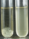
Mutant B. mycoides SIN strains loose aggregation in liquid culture. Wild type strains (left) grow forming aggregated clumps absent in mutants (right).
Figure 4
Figure 4
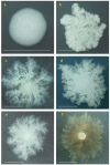
Colony mutants derived from SIN96 strain. Strains were grown on 1.5% agar medium for 50 hours at 25°C. The colony in the center is SIN96, with wild type morphotype and mutants at the periphery: 1) SINett, 2) CAD, 3) CIC, 4–6) cotton-like colonies.
Figure 5
Figure 5
Colony mutants. a) SINett, b) CIC with shorter and thicker trunks, c-d-e-f) cotton like-mutants with different texture and color. Scale bar = 1 cm.
Figure 6
Figure 6
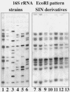
Ribotyping in Bacillus strains. A blot of EcoRI digested total DNA was hybridized by a probe specific for an evolutionarily conserved region of 16S rRNA. Various members of the B. cereus group show different patterns: lane 1, B. mycoides ATCC 6462T; lane 2, B. mycoides DX; lane 3, B. pseudomycoides NRRL B-617T; lane 4, B. mycoides NRRL NRS-273; lane 5, B. thuringiensis BGSC 4D; lane 6, B. mycoides SIN. Mutant strains all derived from SIN96, with identical patterns: lane 7, B. mycoides SINett; lane 8, CAD; lane 9, CIC; lane 10–13 cotton-like mutants.
Figure 7
Figure 7
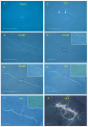
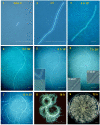
Growth of SIN on agar as seen at the light microscope. Starting from 3 cells (1) the filament grows by division of the cells at both edges (2,3,4,5). The ramification and angle, pointed by arrows in (2), remain at the same distance in the following stages confirming growth at the periphery. In (6) the curvature specific for the strain is apparent at both ends. New filaments start from rupture points, running along the first filament (7) and projecting outside. Insets show enlargements of the region pointed by arrows in panel 2. In (8), advanced stage of colony formation: the structure is enriched by many filaments, becoming as in Fig. 1 after 24 hours. Scale bar: 1–4 = 100μm; 5–8 = 500 μm.
Figure 8
Figure 8
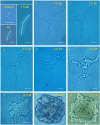
Growth of DX on agar as seen at the light microscope. The filament starting from 3 cells in (1) grows at the periphery (2, 3) and in (4) the specific curvature appears at both ends when the filament is still unique. In (5) the first ramification occurs, enlarged in insets that show duplication and rupture of the filament. In (6) the circle is closed and insets show the rapid cell progression at the closure point. In (7) many lateral ramifications emerge. In (8) these ramifications converge in a few big trunks displaying the specific curvature. In (9) the final shape of the colony is shown. Scale bar: 1–4 = 100 μm; 5–8 = 500 μm; 9 = 1 cm.
Figure 9
Figure 9
Growth of SINett on agar as seen at the light microscope. In this SIN mutant a bent appears at an early stage of colony formation (1) and a double rupture occurs (2). From these ruptures 3 parallel filaments start growing (3). In (4) new ruptures in the upper filament appear. In (5–6) many filaments aggregate in bundles covering the central part of the starting filament. Note that magnification is the same in these 6 figures. In (7) the colony covers almost all of the central space. In (8–9) the final shape is shown. Several layers of cells are superimposed. They appear more evident in (9) where the picture was taken with a filter. Scale bar: 1–6 = 20 μm, 7–9 = 100 μm.
Figure 10
Figure 10
Autolysis of filaments. Magnifications of two details of Fig. 9/7, showing degradation of filaments remaining outside the colony mass.
Figure 11
Figure 11
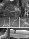
Scanning electron microscopy of wild type SIN colony on agar plate. Bacilli form filaments expanding on agar towards periphery, leaving wide empty spaces (a). Trunks fuse proceeding together, or separate. Details of the same picture show pioneer filaments proceeding independently (b), going in parallel (c), fusing and separating (d). High magnifications (25–30.000 X) of the SIN cells show no twisting of the structures of the external cell wall (e-f).
Figure 12
Figure 12
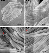
Scanning electron microscopy on agar of the mutant SINett. In (a) the entire colony at 9 hours. At variance with wild type, all bundles turn towards the colony center. (b) shows frequent cell separations along the filaments; (c-d) show the presence of minicells (indicated by arrows).
Figure 13
Figure 13
Sequence conservation. Translated ORFs of a region of the "division cell wall" cluster of B. mycoides SIN are compared to those of other Bacillus cereus group members. Numbers indicate percent of identical amino acids in the ORFs.

Similar articles

Cited by

References

    1. Shapiro JA. The significance of bacterial colony patterns. BioEssays. 1995;17:597–607. - PubMed
    1. Rudner R, Martsinkevich O, Leung W, Jarvis ED. Classification and genetic characterization of pattern forming Bacilli. Molecular Microbiology. 1998;27:687–703. doi: 10.1046/j.1365-2958.1998.00717.x. - DOI - PubMed
    1. Shapiro JA. Thinking about bacterial populations as multicellular organisms. Annu Rev Microbiol. 1998;52:81–104. doi: 10.1146/annurev.micro.52.1.81. - DOI - PubMed
    1. Ben-Jacob E, Levine H. The artistry of nature. Nature. 2001;409:985–986. doi: 10.1038/35059178. - DOI - PubMed
    1. Seo JW, Ohnishi Y, Hirata A, Horinouchi S. ATP-binding cassette transport system involved in regulation of morphological differentiation in response to glucose in Streptomyces griseus. J Bacteriol. 2002;184:91–103. doi: 10.1128/JB.184.1.91-103.2002. - DOI - PMC - PubMed

Publication types

MeSH terms

Associated data

LinkOut - more resources