. 2002 Nov;13(11):3859-69.
doi: 10.1091/mbc.e02-04-0205.

Epsilon-tubulin is an essential component of the centriole

Affiliations

Epsilon-tubulin is an essential component of the centriole

Susan K Dutcher et al. Mol Biol Cell. 2002 Nov.

Abstract

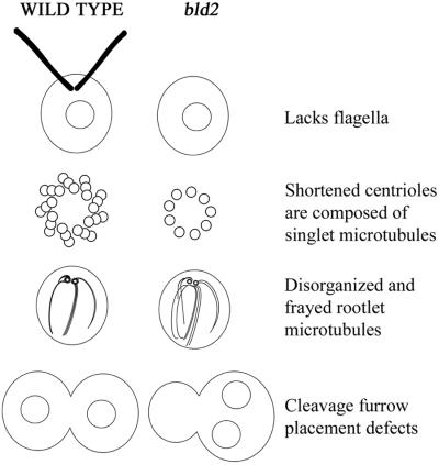
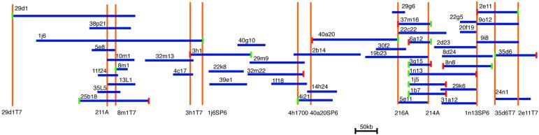
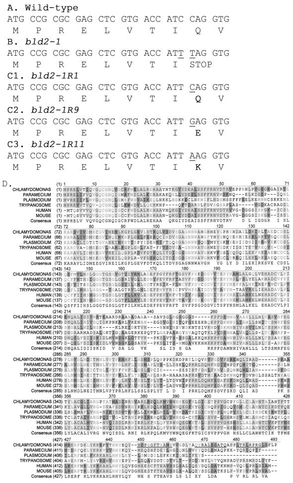
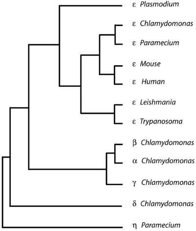
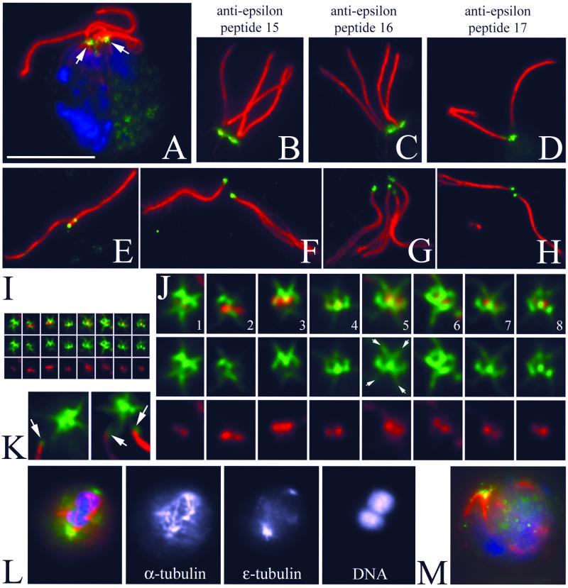
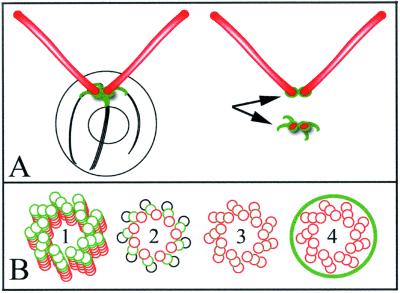
Centrioles and basal bodies are cylinders composed of nine triplet microtubule blades that play essential roles in the centrosome and in flagellar assembly. Chlamydomonas cells with the bld2-1 mutation fail to assemble doublet and triplet microtubules and have defects in cleavage furrow placement and meiosis. Using positional cloning, we have walked 720 kb and identified a 13.2-kb fragment that contains epsilon-tubulin and rescues the Bld2 defects. The bld2-1 allele has a premature stop codon and intragenic revertants replace the stop codon with glutamine, glutamate, or lysine. Polyclonal antibodies to epsilon-tubulin show peripheral labeling of full-length basal bodies and centrioles. Thus, epsilon-tubulin is encoded by the BLD2 allele and epsilon-tubulin plays a role in basal body/centriole morphogenesis.


Figures

Figure 1
Figure 1
Diagram of phenotypes of wild-type and bld2-1 cells. The primary defect in bld2-1 cells is likely to be shortened centrioles with singlet microtubules. The consequences of this defect are failure to assemble flagella, disorganized and frayed rootlet microtubules, and defects in cleavage furrow placement. Not shown are defects in centrin assembly and premature initiation of the cleavage furrow.
Figure 2
Figure 2
Diagram of BAC clones mapped to linkage group III surrounding the BLD2 locus. The total chromosome walk covers 720 kb and 4.5 map units. Dark blue lines represent the BAC clones and their length as determined by fingerprinting gels (Marra et al., 1997). Orange lines represent the position of the probes used in the walk and their names are given below the line. For example, 40a20SP6 represents a probe generated from the sequence on the SP6 side of the BAC 40a20. Small red boxes are the SP6 side of the BAC and small green boxes are on the T7 side of the walk. BACs 11 m13, 1h1, 18k12, 22a5, 7n18, 27c21, 8122, 8f21, 8d23, and 8g22 were found in this region, but were not placed on the map.
Figure 3
Figure 3
Nucleotide and amino acids changes in bld2 alleles and alignment of ε-tubulin from multiple organisms. (A) Nucleotide and protein sequence for the first 30 bp of the coding region of ε-tubulin from wild-type cells. (B) From bld2-1 cells, 5743 bp of sequence were obtained. The nucleotide and protein sequences for the first 30 bp of the coding region are shown. There were two C-to-T transition mutations, which are indicated by underlines. (C) Nucleotide and protein sequence for the first 30 bp of the coding region is shown for three intragenic revertant alleles. In each revertant the TAG stop codon is changed to an amino acid. (D) Protein coding sequence of ε-tubulin predicted using Genie trained on 50 Chlamydomonas genes (Kulp et al., 1996) and aligned with ε-tubulin sequences from human, mouse, Trypanosoma, Plasmodium, and Paramecium (see accession numbers in Figure 4). The peptides used for making antibodies are underlined (MDPGGFTSAME, QYRALEGSTQ, and LTRLRPRG). Light gray indicates conservation of the amino acid in all ε-tubulins, medium gray indicates conservation in >50% of the ε-tubulins, dark gray indicates similarity, and white indicates the lack of conservation.
Figure 4
Figure 4
Phenogram of ε-tubulins. ClustalW was used to align five ε-tubulin sequences, the α, β, γ, and δ-tubulin genes from Chlamydomonas, and η-tubulin from Paramecium, which served as an outgroup for the construction of the tree (Thompson et al., 1994). This alignment was analyzed using the Phylip package (Felsenstein, 1996) to obtain a distance matrix using a PAM matrix and the tree was calculated using neighbor joining. A consensus tree was determined with α-tubulin from Chlamydomonas (A53298), β-tubulin from Chlamydomonas (UBKM), γ-tubulin from Chlamydomonas (AAB71841), δ-tubulin from Chlamydomonas (T07903), ε-tubulin from Chlamydomonas (AF502577), ε-tubulin from human (Q9UJT0), ε-tubulin from mouse (BAB26636), ε-tubulin from Paramecium (CAD20554), ε-tubulin from Plasmodium (AF216743), ε-tubulin from Trypanosoma (AAF32302), and η-tubulin from Paramecium (CAB99490).
Figure 5
Figure 5
Reversion of cleavage furrow placement defect in bld2-1 transformants. Recently divided pairs of daughter cells were photographed and the areas measured. The ratio of the larger to the smaller cell is plotted for 50 cells. (A) Wild-type cells. (B) bld2-1 cells. (C) bld2-1 cells with a BLD2 transgene. Wild-type and bld2-1 cells with a BLD2 transgene produced similar distributions, whereas both were significantly different from the bld2-1 cells.
Figure 6
Figure 6
Immunofluorescence of Chlamydomonas with anti-ε-tubulin peptide antibodies labels an unusual pattern associated with basal bodies and striated fibers. (A) Triple fluorescence of a Chlamydomonas dikaryon: DNA is labeled with 4,6-diamidino-2-phenylindole (blue), the mouse monoclonal 6-11B-1 identifies acetylated α-tubulin (red), and ε-tubulin is labeled with the rabbit polyclonal p15 peptide antibody (green). Bar, 10 μm. Magnification is conserved in the subsequent panels, unless noted. One of the basal body apparatuses is from the wild-type parent and the other is from the rgn1 bld2-1 parent. (B–D) Three independent antipeptide antibodies label the basal bodies in an identical manner. Red is acetylated α-tubulin and green is anti-ε-tubulin p15 (B), p16 (C), or p17 (D). (E–H) ε-Tubulin is associated with the proximal end of flagella detached from Chlamydomonas cell bodies. The staining appears as a discrete spot that is opposite the end of the flagella that has begun to fray (especially note F and G). E and F are labeled with anti-p15 ε-tubulin antibody. G and H are stained with anti-p17 antibody. (I and J) Staining of a basal body associated structure that is left when flagella become detached from the Chlamydomonas cell body. The basal bodies are labeled with acetylated α-tubulin (red) and appear as a pair of dots. The ε-tubulin (green) seems to surround the acetylated α-tubulin dots, creating a figure-eight–shaped cage. Additional staining extends in a “cross-shaped” pattern, reflecting association of ε-tubulin with the striated fibers (arrows in 5). (I) Montage of basal body images shown at the same magnification as the previous panels. (J) Threefold enlargement of this montage. (K) Enlarged image of detached flagella adjacent to the cell body showing the regions of staining associated with the proximal tip of the flagellum (arrows) and retained in the cell body. (L) Merged image of a telophase cell showing ε-tubulin staining (green) at the poles of the spindle (red). Single-channel images with staining of α-tubulin, ε-tubulin, and DNA. (M) bld2-1 cell showing staining with ε-tubulin in the basal body region (green), disorganized rootlet microtubules (red), and no flagella.
Figure 7
Figure 7
Model of ε-tubulin localization and function within the basal body and centriole. (A, left) When intact Chlamydomonas are labeled with the ε-tubulin antibody, small fibrous structures at the base of the flagella are labeled, which reflects association with the basal bodies. (A, top right) A small spot of staining is associated with the proximal end of detached flagella. (A, bottom right) Chlamydomonas cell bodies with flagella detached show labeling of a small distinctive structure consisting of a figure-eight–shaped cage surrounding the two basal body and/or probasal body spots, with four spikes of staining extending along the striated fibers. (B) bld2-1 cells have disrupted triplet microtubules and basal bodies/centrioles have singlet microtubules. This observation, coupled with the cage-like pattern of ε-tubulin staining suggest several models by which ε-tubulin stabilizes triplet microtubules. These include 1) ε-tubulin serving as a plus end cap to the basal body microtubules, 2) ε-tubulin contributing to the B (or B and C) subfiber microtubule lattice as a subunit, 3) ε-tubulin creating the unusual junction of the B (or B and C) subfiber microtubule structure with the A microtubule, or 4) ε-tubulin forming a cage around the entire centriole that confers stability or assembly properties to this structure.

References

    1. Barnes WM. PCR amplification of up to 35 kb DNA with high fidelity and high yield from lambda bacteriophage templates. Proc Natl Acad Sci USA. 1994;91:2216–2220. - PMC - PubMed
    1. Bobinnec Y, Khodjakov A, Mir LM, Rieder CL, Eddé B, Bornens M. Centriole disassembly in vivo and its effects on centrosome structure and function in vertebrate cells. J Cell Biol. 1998;143:1575–1589. - PMC - PubMed
    1. Chang P, Stearns T. Delta-tubulin and epsilon-tubulin: two new human centrosomal tubulins reveal new aspects of centrosome structure and function. Nat Cell Biol. 2000;2:30–35. - PubMed
    1. Cole DG, Diener DR, Himelblau AL, Beech PL, Fuster JC, Rosenbaum JL. Chlamydomonas kinesin-II-dependent intraflagellar transport (IFT): IFT particles contain proteins required for ciliary assembly in Caenorhabditis eleganssensory neurons. J Cell Biol. 1998;141:993–1008. - PMC - PubMed
    1. Dippell RV. The development of basal bodies in Paramecium. Proc Natl Acad Sci USA. 1968;61:461–468. - PMC - PubMed

Publication types

MeSH terms

LinkOut - more resources