. 2002 Nov;13(11):3890-900.
doi: 10.1091/mbc.e02-06-0332.

Meiotic cohesion requires accumulation of ORD on chromosomes before condensation

Affiliations

Meiotic cohesion requires accumulation of ORD on chromosomes before condensation

Eric M Balicky et al. Mol Biol Cell. 2002 Nov.

Erratum in

  • Mol Biol Cell. 2003 Apr;14(4):following 1743

Abstract

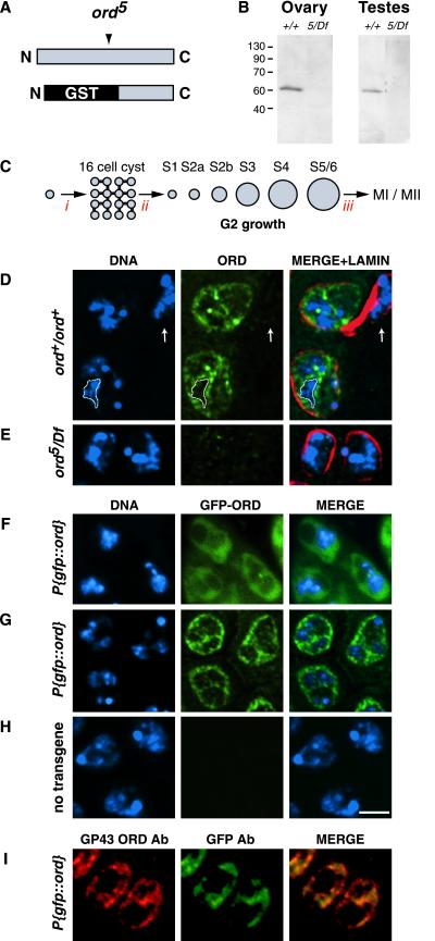
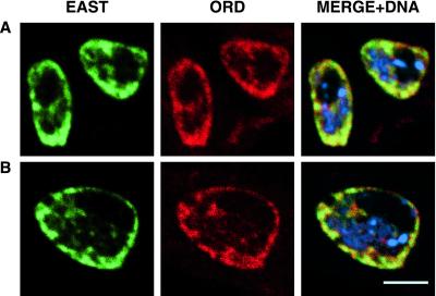
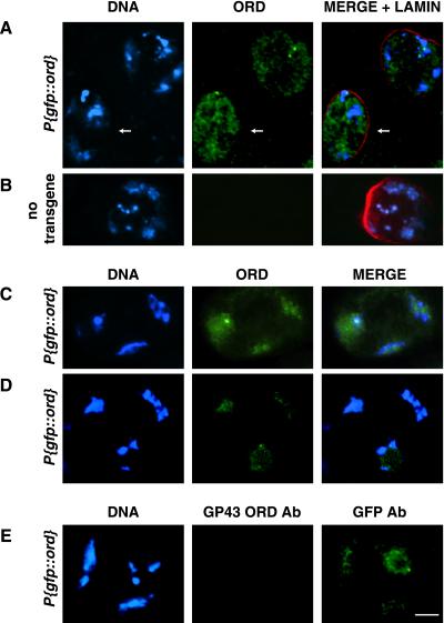
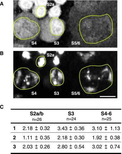
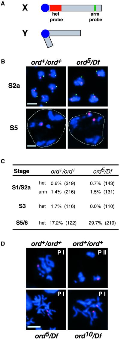
Cohesion between sister chromatids is a prerequisite for accurate chromosome segregation during mitosis and meiosis. To allow chromosome condensation during prophase, the connections that hold sister chromatids together must be maintained but still permit extensive chromatin compaction. In Drosophila, null mutations in the orientation disruptor (ord) gene lead to meiotic nondisjunction in males and females because cohesion is absent by the time that sister kinetochores make stable microtubule attachments. We provide evidence that ORD is concentrated within the extrachromosomal domains of the nuclei of Drosophila primary spermatocytes during early G2, but accumulates on the meiotic chromosomes by mid to late G2. Moreover, using fluorescence in situ hybridization to monitor cohesion directly, we show that cohesion defects first become detectable in ord(null) spermatocytes shortly after the time when wild-type ORD associates with the chromosomes. After condensation, ORD remains bound at the centromeres of wild-type spermatocytes and persists there until centromeric cohesion is released during anaphase II. Our results suggest that association of ORD with meiotic chromosomes during mid to late G2 is required to maintain sister-chromatid cohesion during prophase condensation and that retention of ORD at the centromeres after condensation ensures the maintenance of centromeric cohesion until anaphase II.


Figures

Figure 1
Figure 1
ORD localization in early G2 primary spermatocytes. (A) Schematic of full-length ORD protein showing relative position of the ord5 nonsense mutation and the GST-ORD immunogen (bottom) against which GP43 antibodies were raised. (B) Western blot of Drosophila ovary (15 μg/lane) and testis (4 sets/lane) extracts probed with GP43 ORD antiserum. (C) Spermatocyte development in D. melanogaster (not to scale). A spermatagonial cell undergoes four incomplete mitotic divisions (i) to generate a cyst of 16 primary spermatocytes. After S phase (ii), spermatocytes proceed through an extensive G2 phase that can be divided into seven stages (Cenci et al., 1994). Subsequently, chromosome condensation occurs (iii) and cells undergo meiosis I and II. (D and E) Fixed testes squashes from wild-type and ord5/Df third instar larvae were immunostained using GP43 ORD antiserum. DNA (blue), ORD (green), nuclear lamin (red). Images represent either a single section (D) or full projection (E) of a deconvolved z-series. The extrachromosomal distribution of ORD is emphasized by the outlined region in D, which surrounds a single chromosomal territory. ORD staining is absent in the somatically derived cyst cell (arrow). (F-H) GFP-ORD localization in S2a primary spermatocytes. DNA (blue), GFP-ORD (green). (F) GFP fluorescence in live spermatocytes from a P{gfp::ord} third instar larva. GFP-ORD is primarily nuclear, as determined by phase contrast microscopy (not shown). (G and H) Immunofluorescence detection of GFP-ORD in fixed testes squashes using anti-GFP antibody. (G) P{gfp::ord} transgenic spermatocytes, single section from a deconvolved z-series. (H) Nontransgenic control spermatocytes, full projection of a deconvolved z-series. (I) P{gfp::ord} transgenic spermatocytes costained with GP43 ORD (red) and GFP (green) antibodies. Both antibodies detect the same distribution of ORD protein at this stage. All images are same scale. Bar, 5 μm.
Figure 2
Figure 2
ORD and EAST colocalize in early G2 primary spermatocytes. Fixed testes squashes from wild-type third instar larvae were immunostained with GP43 ORD and EAST polyclonal antisera and subjected to confocal analysis. EAST (green), ORD (red), DNA (blue). (A) S2a spermatocytes. (B) A single S2b spermatocyte. Bar, 5 μm.
Figure 3
Figure 3
ORD accumulates on chromatin in late G2 spermatocytes. Localization of GFP-ORD in spermatocytes from P{gfp::ord} (A, C, D, and E) or nontransgenic (B) males. ORD (green), DNA (blue), nuclear lamin (red). (A) Immunofluorescence detection of GFP-ORD in S4 spermatocytes using GFP antibodies. Nuclear distribution of ORD appears more homogeneous (arrow) than in earlier stages (compare with Figure 1G), and colocalization with chromatin is visible in a slightly older cell (unlabeled, top right). Image is a single section from a deconvolved z-series. (B) GFP immunofluorescence signal is absent in a nontransgenic S4 primary spermatocyte. Image represents a full projection of a nondeconvolved z-series. (C) GFP-ORD signal detected with GFP antibody coincides with DNA in an S5 spermatocyte. (D) GFP fluorescence in a live S6 spermatocyte. (E) Late G2 P{gfp::ord} primary spermatocyte costained with GP43 ORD (red) and GFP (green) antibodies. All images are same scale. Bar, 10 μm.
Figure 4
Figure 4
Quantification of ORD protein in spermatocyte nuclei during G2. (A) Immunofluorescence detection of GFP-ORD and (B) DAPI staining of DNA in P{gfp::ord} spermatocytes. Nuclei are outlined in yellow. Bar, 10 μm. ORD staining appears to diminish during spermatocyte growth. (C) Three-dimensional reconstructions of deconvolved z-series from three different fields of cells were used to calculate the total amount of ORD protein within the nuclei of spermatocytes at different stages of G2 growth. GFP-ORD values were normalized against the average DNA signal for the corresponding stage. The average of the normalized GFP-ORD values is shown for each stage. The number of cells used to calculate the average is indicated below each stage designation. A subset of cells from field 1 is shown above, with specific stages labeled. Units are arbitrary.
Figure 5
Figure 5
Cohesion defects in ordnull spermatocytes become manifest in late G2. (A) Schematic representation of sex chromosomes indicating the X chromosome probes used for FISH analysis. Centromere-proximal probe directed against 359-base pair satellite repeat is shown in red, and the arm probe spanning region 3C1–6 (BACR34O03) is green. (B) Full projection z-series of wild-type and ord5/Df spermatocytes hybridized with het (red) and arm (green) probes. DNA is shown in blue and a white line designates the perimeter of a single nucleus in the S5 spermatocyte panels. In both genotypes, single FISH signals for each probe are apparent during the S2a stage. Two het signals in a single ord5/Df spermatocyte nucleus at stage S5 indicate that sister chromatids have prematurely separated. Arm signal is not visible in older spermatocytes. Bars, 5 μm. (C) Percentage of cells displaying separated sister FISH signals at various stages of G2 growth in wild-type and ord5/Df spermatocytes. Three to four slides were scored for each genotype, and the total number of cells scored is shown in parentheses. (D) The centromere-proximal FISH probe (red) was used to assay cohesion after DNA (blue) condensation. Prophase figures from wild-type (top) as well as ord5/Df and ord10/Df spermatocytes (bottom) are shown. Prophase I and II are designated by PI and PII, respectively. The nucleus of a single spermatocyte is shown in each panel. Bar, 5 μm.
Figure 6
Figure 6
ORD remains at the centromeres of condensed meiotic chromosomes until cohesion is released at anaphase II. Immunolocalization of GFP-ORD on fixed testes squashes from P{gfp::ord} (A-H, and K) or nontransgenic (I and J) adult flies. (A, B, C, E, G, and I) show GFP-ORD (green) and DNA (blue) during meiotic progression. Distinct GFP-ORD foci are visible on condensed chromosomes from prometaphase I until sister chromatids segregate at anaphase II (compare A, B, C, and E, with G). GFP-ORD colocalizes with MEI-S332 (red) at the centromeres (C and E). All images are either partial (A, B, C, and E) or full (G and I) projections of deconvolved z-series and are same scale. Bar, 2 μm. (D, F, H, and J) Corresponding whole cell images showing DNA (blue) and tubulin (red). Arrow in H designates the chromatin mass shown in G. (K) Nucleus of a telophase I P{gfp::ord} transgenic spermatocyte stained with both GP43 ORD (red) and GFP (green) antibodies. Four chromosomal foci are detected by GFP antibodies, but are not recognized by GP43 ORD antibodies. Bars, 5 μm.

References

    1. Amon A. The spindle checkpoint. Curr Opin Genet Dev. 1999;9:69–75. - PubMed
    1. Bickel SE, Moore DP, Lai C, Orr-Weaver TL. Genetic interactions between mei-S332 and ord in the control of sister-chromatid cohesion. Genetics. 1998;150:1467–1476. - PMC - PubMed
    1. Bickel SE, Orr-Weaver TL, Balicky EM. The sister-chromatid cohesion protein ORD is required for chiasma maintenance in Drosophila oocytes. Curr Biol. 2002;12:925–929. - PubMed
    1. Bickel SE, Orr-Weaver TL. Holding chromatids together to ensure they go their separate ways. BioEssays. 1996;18:293–300. - PubMed
    1. Bickel SE, Orr-Weaver TL. Regulation of sister-chromatid cohesion during Drosophila meiosis. In: Zirkin BR, editor. Germ Cell Development, Division, Disruption, and Death. New York: Springer-Verlag; 1998. pp. 37–48.

Publication types

MeSH terms