. 2002 Nov;13(11):3967-75.
doi: 10.1091/mbc.02-05-0069.

Microtubule flux and sliding in mitotic spindles of Drosophila embryos

Affiliations

Microtubule flux and sliding in mitotic spindles of Drosophila embryos

Ingrid Brust-Mascher et al. Mol Biol Cell. 2002 Nov.

Abstract

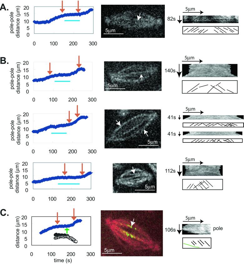
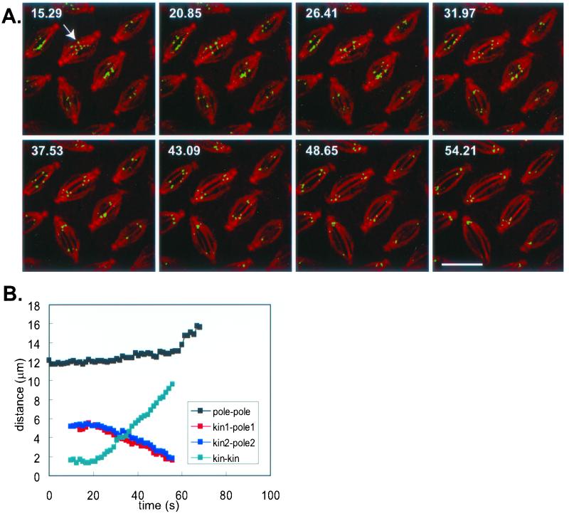
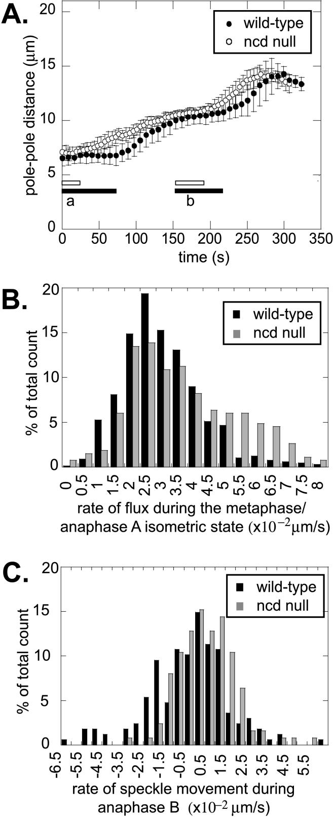
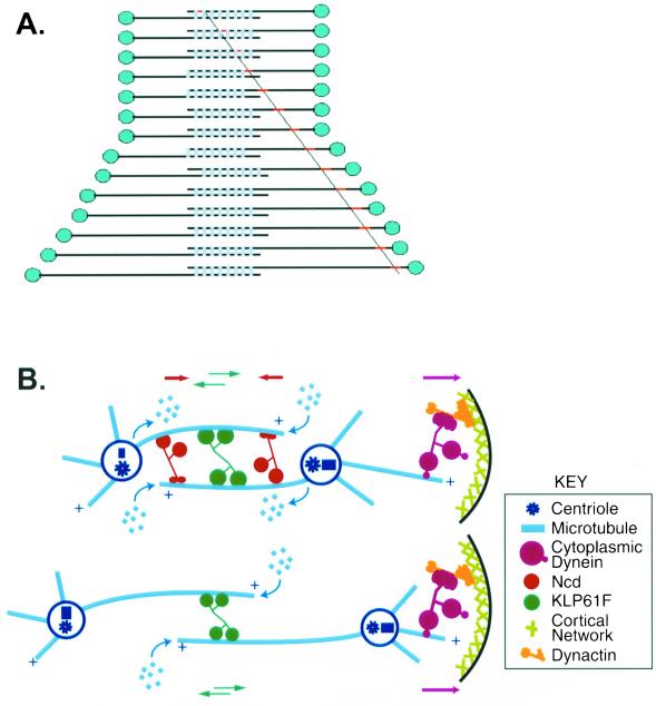
We proposed that spindle morphogenesis in Drosophila embryos involves progression through four transient isometric structures in which a constant spacing of the spindle poles is maintained by a balance of forces generated by multiple microtubule (MT) motors and that tipping this balance drives pole-pole separation. Here we used fluorescent speckle microscopy to evaluate the influence of MT dynamics on the isometric state that persists through metaphase and anaphase A and on pole-pole separation in anaphase B. During metaphase and anaphase A, fluorescent punctae on kinetochore and interpolar MTs flux toward the poles at 0.03 microm/s, too slow to drive chromatid-to-pole motion at 0.11 microm/s, and during anaphase B, fluorescent punctae on interpolar MTs move away from the spindle equator at the same rate as the poles, consistent with MT-MT sliding. Loss of Ncd, a candidate flux motor or brake, did not affect flux in the metaphase/anaphase A isometric state or MT sliding in anaphase B but decreased the duration of the isometric state. Our results suggest that, throughout this isometric state, an outward force exerted on the spindle poles by MT sliding motors is balanced by flux, and that suppression of flux could tip the balance of forces at the onset of anaphase B, allowing MT sliding and polymerization to push the poles apart.


Figures

Figure 1
Figure 1
Speckles flux toward the poles at 0.03 μm/s during the metaphase/anaphase A isometric state, but during anaphase B flux stops and speckles move at the same rate as the poles. Speckle movement was obtained from time lapse confocal images of Drosophila embryos injected with a low concentration of rhodamine labeled tubulin. For each spindle (center panels) we obtained the pole-pole distance as a function of time (graph at left) and followed an individual microtubule bundle to obtain the kymograph for the period of time during which it was clearly visible (shown by red arrows on graph). The horizontal blue lines under the data plots denote the metaphase/anaphase A isometric states. We measured speckle movement using both automatic and manual kymographs (see MATERIALS AND METHODS), which yielded the same results. The kymographs are shown in the right hand panels and the graph below each kymograph shows the trajectories of individual speckles. It should be noted that the printed images are not as clear as the original images analyzed in MetaMorph, from which the data were obtained, because the images were converted from 12 to 8 bits during figure preparation. (A) This kymograph was obtained using MetaMorph's function on a stationary spindle during the metaphase/anaphase A isometric state. (B) These kymographs were obtained manually (see MATERIALS AND METHODS). (C) Manual kymograph for a MT bundle going to a kinetochore. The graph on the left shows the distance between poles (solid blue diamonds) and the distance from two sister kinetochores to the poles (circles). The green arrow shows the onset of anaphase A. In the middle panel red shows tubulin and green shows GFP::CID, a kinetochore marker. The kymograph was obtained for the microtubule bundle going from the pole to the kinetochore (at the equatorial end of the bundle). The green line in the bottom sketch represents the movement of the kinetochore.
Figure 2
Figure 2
Kinetochores move toward the poles at 0.1 μm/s. Kinetochore-to-pole movement was calculated from time lapse confocal images of Drosophila embryos expressing GFP::CID injected with rhodamine tubulin (top). Time is given in seconds in each frame; scale bar, 10 μm. Bottom: the distance between the poles, between two sister kinetochores, and between a kinetochore and the corresponding pole were calculated from the time lapse images (shown here for the spindle marked by the arrow). Anaphase B begins after anaphase A is almost complete. GFP::CID is detectable from prometaphase to anaphase B.
Figure 3
Figure 3
Flux and sliding in Ncd null mutant embryos. (A) Pole-pole distance as a function of time for wild-type (●) and Ncd null (○) embryos during cycle 12. The bars under the data plots represent the prometaphase isometric state (a) and the metaphase/anaphase A isometric state (b) for wild-type (closed bars) and Ncd null (open bars) embryos. In the Ncd mutants, isometric states are shortened and mitosis proceeds faster. (B) Histogram for the rate of flux during the metaphase/anaphase A isometric state for wild-type (black bars) and Ncd null (gray bars) embryos. The number of counts was normalized to the total number (667 for wild-type and 267 for Ncd null). The average is not significantly different. (C) Histogram for the rate of speckle movement during anaphase B for wild-type (black bars) and Ncd null (gray bars) embryos. The number of counts was normalized to the total number (168 for wild-type and 125 for Ncd null). Both are centered around 0, consistent with MT-MT sliding during anaphase B.
Figure 4
Figure 4
Flux is converted to sliding at the onset of anaphase B. (A) Behavior of speckles on ipMTs. During the metaphase/anaphase A isometric state, flux (red dot tracked by diagonal line) is due to subunit addition at plus ends of antiparallel MTs within ipMT bundles (over-lapping horizontal lines) and subunit loss at minus ends coupled to the sliding apart of ipMTs driven by MT sliding motors (light blue bars) so the poles (blue dots) maintain a constant spacing. At anaphase B onset, subunit loss at the poles stops while subunit addition at plus ends and MT-MT sliding continue. This converts flux to net sliding and consequently the poles are pushed apart. (B) Detailed schematic of the metaphase/anaphase A spindle (top) and anaphase B spindle (bottom), showing the three motors previously shown to be involved in mitosis (Sharp et al., 2000a) and sites of tubulin subunit addition and loss within ipMT bundles. During the metaphase/anaphase A isometric state, the action of sliding motors coupled to flux generates the balance of forces that maintains a constant spacing between the spindle poles. Tipping this balance by suppression of depolymerization at the poles leads to anaphase B, which may depend on KLP61F in the spindle interzone driving MT-MT sliding coupled to MT polymerization at the spindle equator (with cortical dynein augmenting pole-to-pole separation late in anaphase B; Sharp et al., 2000a).

References

    1. Cole DG, Saxton WM, Sheehan KB, Scholey JM. A “slow” homotetrameric kinesin-related motor protein purified from Drosophila embryos. J Biol Chem. 1994;269:22913–22916. - PMC - PubMed
    1. Desai A, Maddox PS, Mitchison TJ, Salmon ED. Anaphase A chromosome movement and poleward spindle microtubule flux occur at similar rates in Xenopus extract spindles. J Cell Biol. 1998;141:703–713. - PMC - PubMed
    1. Endow SA, Chandra R, Komma DJ, Yamamoto AH, Salmon ED. Mutants of the Drosophila Ncd microtubule motor protein cause centrosomal and spindle pole defects in mitosis. J Cell Sci. 1994a;107:859–867. - PubMed
    1. Endow SA, Kang SJ, Satterwhite LL, Rose MD, Skeen VP, Salmon ED. Yeast Kar3 is a minus-end microtubule motor protein that destabilizes microtubules preferentially at the minus ends. EMBO J. 1994b;13:2708–2713. - PMC - PubMed
    1. Endow SA, Komma DJ. Centrosome and spindle function of the Drosophila Ncd motor protein visualized in live embryos using Ncd-GFP fusion proteins. J Cell Sci. 1996;109:2429–2442. - PubMed

Publication types

LinkOut - more resources