. 2002 Nov;13(11):4045-59.
doi: 10.1091/mbc.e02-05-0294.

Motility determinants in WASP family proteins

Affiliations

Motility determinants in WASP family proteins

Defne Yarar et al. Mol Biol Cell. 2002 Nov.

Abstract

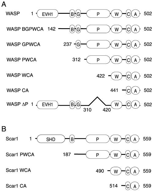
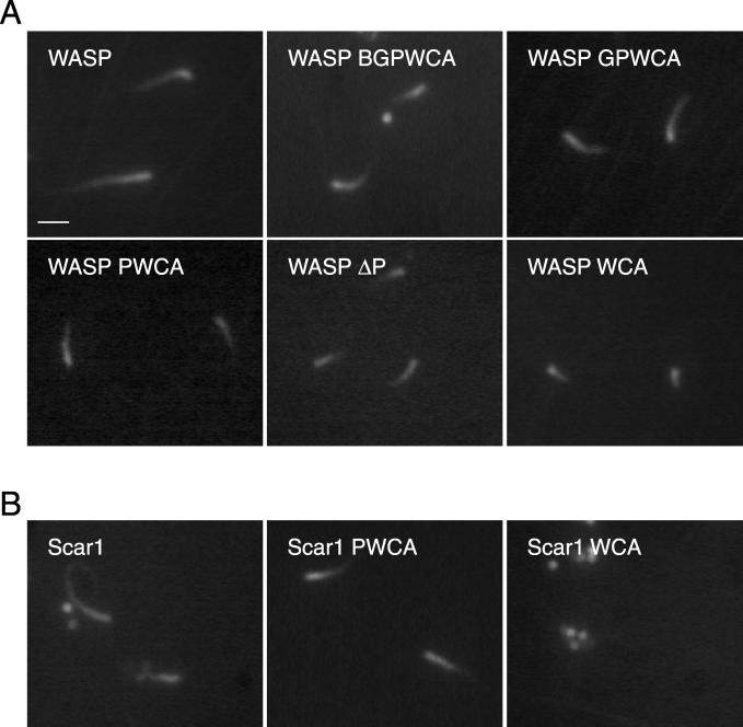
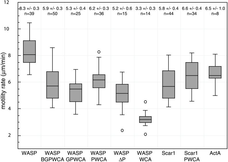
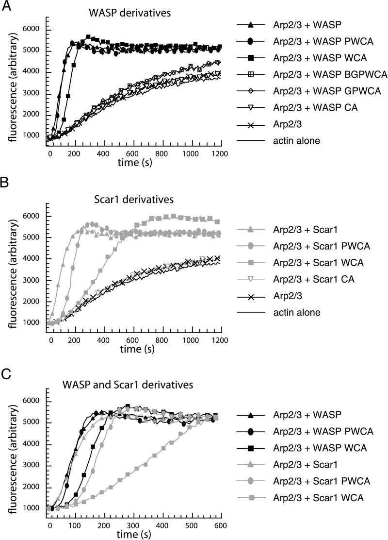
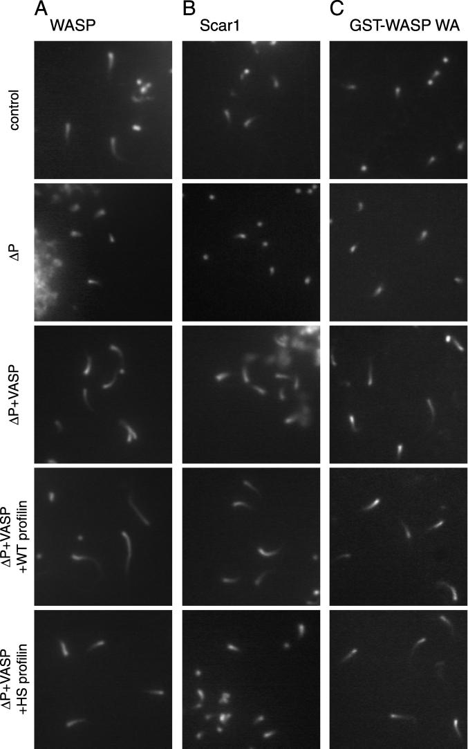
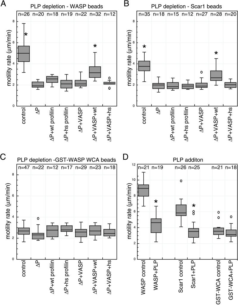
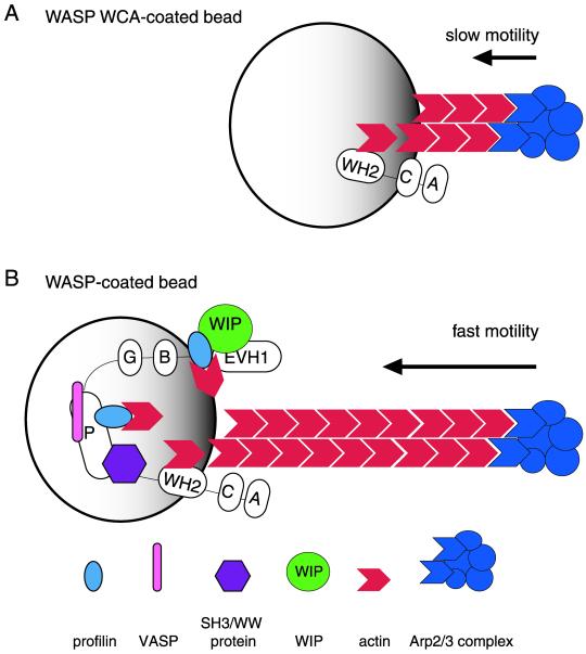
In response to upstream signals, proteins in the Wiskott-Aldrich Syndrome protein (WASP) family regulate actin nucleation via the Arp2/3 complex. Despite intensive study of the function of WASP family proteins in nucleation, it is not yet understood how their distinct structural organization contributes to actin-based motility. Herein, we analyzed the activities of WASP and Scar1 truncation derivatives by using a bead-based motility assay. The minimal region of WASP sufficient to direct movement was the C-terminal WCA fragment, whereas the corresponding region of Scar1 was insufficient. In addition, the proline-rich regions of WASP and Scar1 and the Ena/VASP homology 1 (EVH1) domain of WASP independently enhanced motility rates. The contributions of these regions to motility could not be accounted for by their direct effects on actin nucleation with the Arp2/3 complex, suggesting that they stimulate motility by recruiting additional factors. We have identified profilin as one such factor. WASP- and Scar1-coated bead motility rates were significantly reduced by depletion of profilin and VASP and could be more efficiently rescued by a combination of VASP and wild-type profilin than by VASP and a mutant profilin that cannot bind proline-rich sequences. Moreover, motility of WASP WCA beads was not affected by the depletion or addback of VASP and profilin. Our results suggest that recruitment of factors, including profilin, by the proline-rich regions of WASP and Scar1 and the EVH1 domain of WASP stimulates cellular actin-based motility.


Figures

Figure 1
Figure 1
N-terminal WASP and Scar1 truncation derivatives. (A) Schematic diagrams of WASP truncation and deletion derivatives. (B) Schematic diagrams of Scar1 truncation derivatives.
Figure 2
Figure 2
Behavior of WASP/Scar1 derivative-coated beads in X. laevis egg extract, visualized by fluorescence microscopy. (A) Actin structures formed by WASP derivative-coated beads. (B) Actin structures formed by Scar1 derivative-coated beads. Images were taken after samples were incubated on the slide for 5 min. Bar, 2 μm.
Figure 3
Figure 3
Box plot distribution of motility rates of WASP/Scar1/ActA-coated beads in X. laevis egg extract. The average rates (± 2× SE; 95% confidence interval) and number of beads in each data set are presented above each distribution. The top line of each box represents the 3rd quartile, the middle line the median, and the bottom line the 1st quartile. The top and bottom whiskers indicate the maximum and minimum rates measured and the circles indicate outliers.
Figure 4
Figure 4
Effects of WASP/Scar1 truncations and the Arp2/3 complex on actin polymerization kinetics, measured using the pyrene-actin polymerization assay. (A) Actin (2 μM) in the presence of 10 nM Arp2/3 complex and 20 nM WASP derivatives. (B) Actin (2 μM) in the presence of 10 nM Arp2/3 complex and 20 nM Scar1 derivatives. (C) Comparison of a subset of WASP and Scar1 derivative curves in A and B.
Figure 5
Figure 5
Effect of PLP depletion on WASP-, Scar1-, and GST-WASP WA bead motility. (A–C) Composite images of actin structures formed around WASP- (A), Scar1- (B), and GST-WASP WCA (C)-coated beads. Panels from top to bottom are: control depleted extract (control), poly-l-proline depleted extract (ΔP), ΔP extract supplemented with 0.2 μM VASP (Δp + VASP), ΔP extract supplemented with 0.2 μM VASP and 2 μM wild-type profilin (Δprofilin + VASP + wt profilin), and ΔP extract supplemented with 0.2 μM VASP and 2 μM H133S profilin (Δprofilin + VASP + HS profilin). Images were taken after samples were incubated on the slide for 30 min. Bar, 2 μm.
Figure 6
Figure 6
Effect of PLP depletion/addition on WASP-, Scar1-, and GST-WASP WCA bead motility. (A–C) Box plot distribution of motility rates of WASP- (A), Scar1- (B), GST-WASP-WCA (C)-coated beads in conditions indicated: control depleted extract (control), poly-l-proline depleted extract (ΔP), ΔP extract supplemented with 0.2 μM VASP and/or 2 μM wild-type profilin (wt), and/or 2 μM H133S profilin (hs). Asterisks (*) denote population of beads that are significantly faster than beads moving in poly-l-proline–depleted extract. (D) Box plot distribution of motility rates of WASP/Scar1/GST-WASP WCA-coated beads in extract supplemented with 10 μM PLP or buffer (control). The number of beads in each data set is presented above each distribution. Asterisks (*) denote population of beads that move at rates significantly different than beads moving in control experiment.
Figure 7
Figure 7
Model explaining the contributions of WASP/Scar1 regions to actin-based motility. (A) WCA derivative of WASP stimulates the nucleation activity of the Arp2/3 complex and is the minimal region that is sufficient to promote actin-based motility. (B) In full-length WASP, the EVH1 domain and P region recruit cytosolic factors, including profilin, that increase the rate of motility by enhancing actin nucleation with the Arp2/3 complex and/or facilitating actin polymerization.

References

    1. Anton IM, Lu W, Mayer BJ, Ramesh N, Geha RS. The Wiskott-Aldrich syndrome protein-interacting protein (WIP) binds to the adaptor protein Nck. J Biol Chem. 1998;273:20992–20995. - PubMed
    1. Bachmann C, Fischer L, Walter U, Reinhard M. The EVH2 domain of the vasodilator-stimulated phosphoprotein mediates tetramerization, F-actin binding, and actin bundle formation. J Biol Chem. 1999;274:23549–23557. - PubMed
    1. Bear JE, Krause M, Gertler FB. Regulating cellular actin assembly. Curr Opin Cell Biol. 2001;13:158–166. - PubMed
    1. Bear JE, Svitkina TM, Krause M, Schafer DA, Loureiro JJ, Strasser GA, Maly IM, Chaga OY, Cooper JA, Borisy GG, Gertler FB. Antagonism between Ena/VASP proteins and actin filament capping regulates fibroblast motility. Cell. 2002;109:509–524. - PubMed
    1. Bernheim-Groswasser A, Wiesner S, Golsteyn RM, Carlier MF, Sykes C. The dynamics of actin-based motility depend on surface parameters. Nature. 2002;417:308–311. - PubMed

Publication types

MeSH terms

LinkOut - more resources