. 2002 Nov;13(11):4088-99.
doi: 10.1091/mbc.02-06-0092.

The diaphanous-related formin mDia1 controls serum response factor activity through its effects on actin polymerization

Affiliations

The diaphanous-related formin mDia1 controls serum response factor activity through its effects on actin polymerization

John W Copeland et al. Mol Biol Cell. 2002 Nov.

Abstract

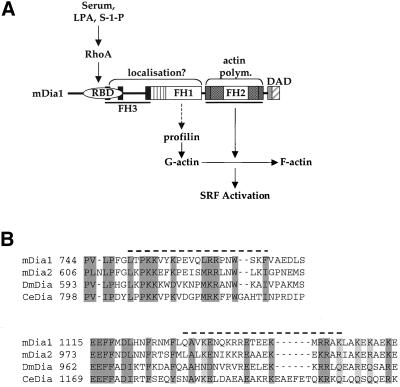
SRF-dependent transcription is regulated by the small GTPase RhoA via its effects on actin dynamics. The diaphanous-related formin (DRF) proteins have been identified as candidate RhoA effectors mediating signaling to SRF. Here we investigate the relationship between SRF activation and actin polymerization by the DRF mDia1. We show that the ability of mDia1 to potentiate SRF activity is strictly correlated with its ability to promote F-actin assembly. Both processes can occur independently of the mDia1 FH1 domain but require sequences in an extended C-terminal region encompassing the conserved FH2 domain. mDia-mediated SRF activation, but not F-actin assembly, can be blocked by a nonpolymerizable actin mutant, placing actin downstream of mDia in the signal pathway. The SRF activation assay was used to identify inactive mDia1 derivatives that inhibit serum- and LPA-induced signaling to SRF. We show that these interfering mutants also block F-actin assembly, whether induced by mDia proteins or extracellular signals. These results identify novel functional elements of mDia1 and show that it regulates SRF activity by inducing depletion of the cellular pool of G-actin.


Figures

Figure 1
Figure 1
Activation of SRF by mDia1 does not require the FH1 domain. (A) Activity of mDia1 deletion constructs. The Rho binding domain (RBD), Formin Homology (FH) domains 1 and 2 (FH1 and FH2) domains, and the three FH3 motifs are indicated. A coiled-coil region homologous to the tail domain of myosin heavy chain (residues 449–550) is shown as a striped box. The minimal DAD domain (residues 1172–1255), predicted by functional analysis of mDia2, is shown as a striped box with a bar indicating the conserved core residues. Expression plasmids for each mDia1 deletion (0.1 μg/dish) were tested in the SRF reporter assay. Inset: relative expression of each protein determined by anti-Flag immunoblots (lines indicate 250, 75-kDa markers in panels 1 and 3, and 50, 25-kDa markers in panel 2). Reporter gene activity is expressed relative to activation by 0.1 μg of SRF-VP16 (see MATERIALS AND METHODS). Results are the mean ± SEM of three independent experiments. (B) SRF activation by the mDia1 FH2 region. Reporter activation by 0.1 μg of FH1/FH2 expression plasmid was compared with that by increasing amounts of expression plasmid encoding the mDia1 FH2 region (FH2; 0.1, 0.3, 1.0 μg). Inset: protein quantitation by anti-9E10 immunoblot. Results are the mean ± SEM of three independent experiments. (C) mDia1 activation of SRF is independent of Rho activity. Reporter activation by the indicated mDia1 derivatives was tested in the presence or absence of expression of the Rho-inactivating C3 transferase. Results are the mean ± SEM of three independent experiments. Relative to the activity observed in its absence, C3 expression reduced activity of the SRF reporter as follows: SRF-VP16, 91.4 ± 5.9%; ΔRBD2, 93 ± 5.7%; FH1FH2, 79.7 ± 12.0%; ΔRBDΔFH1, 87.0 ± 16.5%; FH2, 30.6 ± 1.1%; in parallel experiments C3 expression reduced RhoA. V14-induced SRF activity to 2.4 ± 1.3% (unpublished data).
Figure 2
Figure 2
mDia1 C-terminal sequences required for SRF activation. (A) Deletions within the FH2 region. Expression plasmids encoding N- or C-terminal modifications of the FH2 region were transfected into NIH3T3 cells (0.3 μg/dish) and assayed as in Figure 1 for SRF activation. Proteins are shown schematically at the bottom of each panel, with sequences essential for activity indicated by the boxes. Results are the mean ± SEM of three independent experiments. Inset: relative protein expression (anti-Flag). (B) The extremities of the FH2 region are required in the presence of FH1. Expression plasmids encoding derivatives of FH1/FH2 lacking segments of the FH2 region were transfected into NIH3T3 cells (0.1 μg/dish) and assayed as in Figure 1 for SRF activation. Results are the mean ± SEM of three independent experiments. Inset: relative protein expression (anti-9E10).
Figure 3
Figure 3
mDia1 mutants that activate SRF induce F-actin accumulation. (A) Active mDia1 mutants reorganize cellular F-actin structures. NIH3T3 cells were transfected with plasmids expressing active mDia1 derivatives (FH1/FH2, 0.1 μg; FH2, 1.0 μg) and C3 transferase (0.1 μg) as indicated. F-actin was visualized with FITC-phalloidin. The mDia proteins are shown schematically at the top right, with the essential FH2 sequences shown as boxes. Transfected cells were detected by mDia1 derivative epitope tag immunofluorescence. (B) Inactive mDia proteins do not reorganize cellular F-actin structures. NIH3T3 cells were transfected with plasmids expressing inactive mDia1 derivatives F1F2Δ1, F1F2Δ2, and F1F2ΔC2 (1.0 μg each) as indicated. F-actin was visualized with FITC-phalloidin. Transfected cells were detected by mDia1 derivative epitope tag immunofluorescence. (C) Active mDia1 proteins decrease G: F-actin ratio. NIH3T3 cells were transfected with plasmids expressing FH1/FH2 (0.1 μg), or FH2, F1F2Δ1, F1F2Δ2, and F1F2ΔC2 (1 μg) together with an expression plasmid encoding Flag-tagged wild-type β-actin (0.5 μg). Detergent-soluble and insoluble cell extract fractions were prepared as in MATERIALS AND METHODS, and the amount of Flag-actin in each evaluated by SDS-PAGE and immunoblotting with M2 anti-Flag antibodies. SwinholideA and jasplakinolide treated controls are shown in the bottom panel. A representative experiment is shown (N = 3). (D) SRF activation correlates with ability to promote F-actin assembly. NIH 3T3 cells were transfected with plasmids expressing the indicated mDia1 derivatives as above and the mean F-actin content of transfected cells determined, relative to untransfected cells in the same population, using the FACS. Data represent the mean ± SEM of three independent experiments.
Figure 4
Figure 4
Actin acts downstream of mDia in SRF activation. (A) Activation of SRF is inhibited by nonpolymerizable actin. NIH3T3 cells were transfected with SRF reporter and either 0.1 μg of expression plasmid encoding mDia1 FH1/FH2, together with increasing amounts (0.1, 0.3, 1.0 μg) of plasmids expressing wild-type β-actin or the nonpolymerizable actin G13R, which contains a mutation in the ATP binding cleft; or 0.1 μg plasmid expressing the constitutively active SRF derivative SRF-VP16 with 1.0 μg of each actin expression plasmid. Reporter activation is presented as mean ± SEM of three independent experiments. (B) Expression of wild-type or nonpolymerizable actin does not inhibit mDia-induced F-actin accumulation. NIH 3T3 cells were transfected with expression plasmids encoding FH1/FH2 (0.1 μg) and either wild-type actin or actin G13R (1.0 μg each). Mean F-actin content of transfected cells was quantified relative to that of untransfected cells in the same population using the FACS. Results are the mean ± halfrange of two independent experiments. Wild-type actin expression alone increased mean cellular F-actin levels by up to 40%, whereas expression of G13R alone has no effect on mean cellular F-actin content (Posern et al., 2002). (C) SRF activation by Src. NIH3T3 cells were transfected with SRF reporter and either 0.1 μg of expression plasmid encoding Src Y527F, together with plasmids expressing either C3 transferase (0.1 μg), wild-type or G13R actin (1.0 μg each). Reporter activation is presented as mean ± SEM of three independent experiments.
Figure 5
Figure 5
Interfering mutants of mDia1. (A) The inactive mDia1 mutants F1F2Δ1, F1F2Δ2, and F1F2ΔC2 inhibit SRF activation by activated mDia1. NIH3T3 cells were transfected with SRF reporter and plasmids expressing F1F2Δ1, F1F2Δ2, or F1F2ΔC2 (2.0 μg), together with activated mDia1 (FH1/FH2, 0.1 μg or FH2, 0.5 μg) or SRF-VP16 (0.1 μg). Reporter activation is presented as mean ± SEM of three independent experiments. (B) Activated ΔGBD-Dia2 is inhibited by mDia1 interfering mutants. NIH3T3 cells were transfected with SRF reporter and plasmids expressing F1F2Δ1, F1F2Δ2, F1F2ΔC2, FH1, or F2ΔN2 (2.0 μg each), together with ΔGBD-Dia2 (0.1 μg). Reporter activation is presented as mean ± SEM of three independent experiments. Interfering mDia1 mutants and ΔGBD-Dia2 are shown schematically at the right.
Figure 6
Figure 6
mDia1 interfering derivative F1F2Δ1 prevents reorganization of the F-actin cytoskeleton by activated mDia proteins. (A) NIH3T3 cells were transfected with plasmids expressing either the activated FH1/FH2 mDia1 derivative (0.1 μg; top panels) or the activated mDia2 derivative ΔGBD-Dia2 (bottom panels), together with a plasmid expressing F1F2Δ1 (2.0 μg) as indicated. F-actin was visualized with FITC-phalloidin. (B) NIH3T3 cells were transfected with expression plasmids encoding mDia1 and mDia2 derivatives as in A. Mean F-actin content of transfected cells was quantified relative to that of untransfected cells in the same population using FACS. Results are the mean ± SEM of three independent experiments. The inhibition by F1F2Δ1 is significant (p < 0.05) by Student's t test.
Figure 7
Figure 7
mDia1 interfering derivatives block SRF activation and cytoskeletal rearrangements. (A) Interfering derivatives block activation of SRF by serum stimulation. NIH3T3 cells were transfected with SRF reporter and plasmids expressing mDia1 derivatives F1F2Δ1, F1F2Δ2, F1F2ΔC2, or FH3/M (2.0 μg), or wild-type β-actin (1.0 μg), maintained in 0.5% FCS for 36 h, and then stimulated with 15% serum as indicated. Reporter activation is presented as mean ± SEM of three independent experiments. (B) F1F2Δ1 does not block activation of TCF Elk-1 by serum stimulation. NIH3T3 cells were transfected with the LexA operator controlled reporter LexOP-Luc (0.1 μg), an expression plasmids encoding NLex. ElkC (0.1 μg), a chimeric transcription factor comprising the C-terminal activation domain of Elk-1 fused to the bacterial LexA repressor (Marais et al., 1993), and mDia1 derivative F1F2Δ1 (2.0 μg). Cells were serum-stimulated as in A. Reporter activation is presented as mean ± halfrange of two independent experiments. (C) F1F2Δ1 blocks activation of SRF by LPA stimulation. NIH3T3 cells were transfected with SRF reporter and plasmids expressing mDia1 derivative F1F2Δ1 (2.0 μg), or wild-type β-actin (1.0 μg), maintained in 0.5% FCS for 36 h, and then stimulated with 10 μM LPA as indicated. Reporter activation is presented as mean ± SEM of three independent experiments. (D) Src activity is not required for serum-stimulated SRF activation. NIH3T3 cells were transfected with SRF reporter and plasmids expressing C-terminal Src Kinase (CSK; 2.0 μg) or kinase inactive SrcK298 M (SrcKD; 2.0 μg) and processed as in A. Where indicated, cells were pretreated for 1 h before stimulation with the Src inhibitor PP2 (0.25 μM, 1.0 μM). Reporter activation is presented as mean ± SEM of three independent experiments. (E) F1F2Δ1 blocks stress fiber induction by LPA. NIH3T3 cells were transfected with a plasmid expressing mDia1 derivative F1F2Δ1 (2.0 μg), maintained in 0.5% FCS for 36 h, and then stimulated with 10 μM LPA for 2 min before fixation and staining for F-actin and the transfected protein epitope tag. Arrows indicate the transfected cells.
Figure 8
Figure 8
Functional domains in mDia1. (A) Speculative model for mDia1 functional domains. mDia1 is activated by Rho-GTP binding to the RBD, relieving interaction with the DAD and exposing the FH1 and FH2 domains (Watanabe et al., 1999; Tominaga et al., 2000). The figure shows functional regions within mDia1 as deduced from the studies of SRF activation and actin polymerization. The FH2 region, C-terminal to the FH1 and excluding the DAD homology region is sufficient to induce actin polymerization and thereby activate SRF by a mechanism involving depletion of the G-actin pool. The FH1 domain enhances this, possibly by its interaction with profilin or Src. The FH3 and myosin-tail domains, in addition to the FH1 domain, are likely involved in subcellular localization of mDia1. (B) The FH2 region N- and C-termini. Alignment with other Diaphanous Related Formins of the regions deleted from the N- and C-termini of the FH2 region in the interfering mutants F1F2Δ1 (top panel; mDia1 amino acids 750–770) and F1F2ΔC2 (mDia1 amino acids 1130–1150).

References

    1. Alberts AS. Identification of a carboxyl-terminal diaphanous-related formin homology protein autoregulatory domain. J Biol Chem. 2001;276:2824–2830. - PubMed
    1. Arsenian S, Weinhold B, Oelgeschlager M, Ruther U, Nordheim A. Serum response factor is essential for mesoderm formation during mouse embryogenesis. EMBO J. 1998;17:6289–6299. - PMC - PubMed
    1. Bleul CC, Fuhlbrigge RC, Casasnovas JM, Aiuti A, Springer TA. A highly efficacious lymphocyte chemoattractant, stromal cell-derived factor 1 (SDF-1) J Exp Med. 1996;184:1101–1109. - PMC - PubMed
    1. Burger JA, Burger M, Kipps TJ. Chronic lymphocytic leukemia B cells express functional CXCR4 chemokine receptors that mediate spontaneous migration beneath bone marrow stromal cells. Blood. 1999;94:3658–3667. - PubMed
    1. Castrillon DH, Wasserman SA. Diaphanous is required for cytokinesis in Drosophila and shares domains of similarity with the products of the limb deformity gene. Development. 1994;120:3367–3377. - PubMed

Publication types

LinkOut - more resources