

Review
doi: 10.1046/j.1469-7580.2002.00097.x.
Airway and blood vessel interaction during lung development
Affiliations
- PMID: 12430957
- PMCID: PMC1570917
- DOI: 10.1046/j.1469-7580.2002.00097.x
Item in Clipboard
Review
Airway and blood vessel interaction during lung development
J Anat.
2002 Oct.
Abstract
In the adult lung the pulmonary arteries run alongside the airways and the pulmonary veins show a similar branching pattern to the arteries, though separated from them. During early fetal development the airways act as a template for pulmonary blood vessel development in that the vessels form by vasculogenesis around the branching airways. In later lung development the capillary bed is essential for alveolar formation. This paper reviews evidence for the interaction of the airways and blood vessels in both normal and abnormal lung development.
Figures
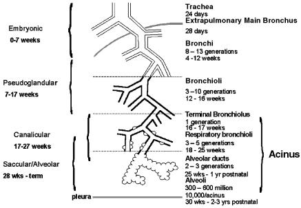
Diagram illustrating the airway structure in the lung and the time period in which each develops.
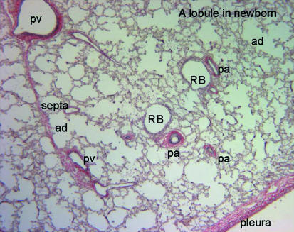
Photomicrograph of a section through a newborn lung stained with elastic van Geison stain to show the relationship of airways and blood vessels. pv, pulmonary vein; pa, pulmonary artery; RB, respiratory bronchiolus; ad, alveolar duct.
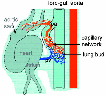
Diagram produced from a serial reconstruction of a 34-day human embryo. The trachea extends from the foregut and the first branches form the lung but on either side of the gut. Around each lung bud is a capillary network which connects cranially to the aortic sac of the heart and caudally to the prospective left atrium. All structures are enveloped in a continuous mesenchymal surround. (Reconstruction by Dr Susan Hall.)
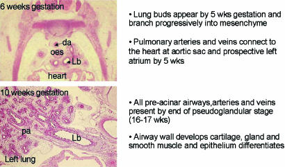
Appearance and summary of growth of the lung in the embryonic and pseudoglandular stage. da, dorsal aorta; oes, oesophagus; Lb, left main bronchus; pa, pulmonary artery.
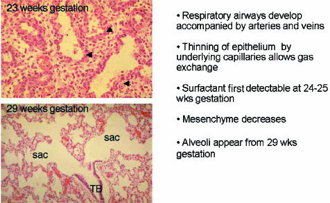
Appearance and summary of growth of the lung in the canalicular and saccular stage. TB, terminal bronchiolus; sac, saccule; arrows, capillaries.
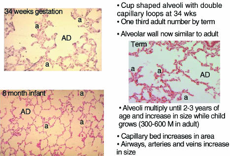
Appearance and summary of growth of the lung in the alveolar stage. AD, alveolar duct; a, alveolus.
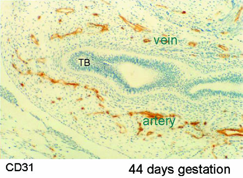
Photomicrograph of section through lung bud at 44 days of gestation. The airway, lined by epithelium (ep), ends in a terminal bud (TB). Around the airway are a large number of capillaries staining positively (brown) for CD31 which recognizes endothelium. On one side of the airway these coalesce to form an artery and on the other side a vein.
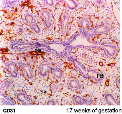
Photomicrograph of lung at 17 weeks of gestation. Arteries accompany airways and veins are found between airways. All vessels stain positively for CD31, the endothelial marker. TB, terminal bud; pa, pulmonary artery; pv, pulmonary vein.
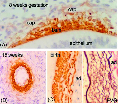
Derivation of smooth muscle cells. (A) Section of airway from a fetus at 8 weeks of gestation, immunostained with α-actin. The bronchial smooth muscle (bsm) stains positively and cells can be seen between this muscle layer and the capillaries (cap) in the mesenchyme. (B) Section of a hilar artery in a fetus of 15 weeks gestation stained for α-actin. The inner layer of cells are strongly labelled and square in shape. Outside are fusiform cells staining weakly for α-actin. (C) A part of a hilar artery stained for α-actin and for elastic van Geison (EVG). The inner smooth muscle cells are a different shape from those in the outer layers though all are within the media. ad, adventitia.
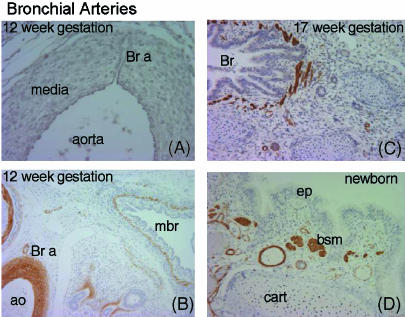
Development of bronchial arteries. (A) Section through dorsal aorta with a bronchial artery (Br a) passing through the media. (B) Hilar region stained with α-actin. The bronchial artery (Br a) can be seen leading from the aorta (ao) to the wall of the main bronchus (mbr). (C) At 17 weeks of gestation many arteries can be seen in the wall of the bronchus. (D) In the newborn the bronchial vessels have increased in size. bsm, bronchial smooth muscle; cart, cartilage; ep, epithelium.
References
-
- Acarregui MJ, Penisten ST, Goss KL, Ramirez K, Snyder JM. Vascular endothelial growth factor gene expresion in human fetal lung in vitro. Am. J. Respir. Cell. Mol. Biol. 1999;20:14–23. - PubMed
-
- Adamson IY, Young L. Alveolar type II cell growth on a pulmonary endothelial extracellular matrix. Am. J. Physiol. 1996;270:L1017–L1022. - PubMed
-
- Auten RL, Jr, Mason SN, Tanaka DT, Welty-Wolf K, Whorton MH. Anti-neutrophil chemokine preserves alveolar development in hyperoxia-exposed newborn rats. Am. J. Physiol. 2001;281:L336–L344. - PubMed
-
- Bhatt AJ, Amin SB, Chess PR, Watkins RH, Maniscalco WM. Expression of vascular endothelial growth factor and Flk-1 in developing and glucocorticoid-treated mouse lung. Pediatr. Res. 2000;47:606–613. - PubMed
-
- Bhatt AJ, Pryhuber GS, Huyck H, Watkins RH, Metlay LA, Maniscalco WM. Disrupted pulmonary vasculature and decreased vascular endothelial growth factor, Flt-1 and Tie-2 in human infants dying with bronchopulmonary dysplasia. Am. J. Respir. Crit. Care Med. 2001;164:1971–1980. - PubMed
Publication types
MeSH terms
LinkOut - more resources
Full Text Sources
