. 2002 Nov 26;99(24):15462-7.
doi: 10.1073/pnas.242328999. Epub 2002 Nov 13.

Role of the thyroid-stimulating hormone receptor signaling in development and differentiation of the thyroid gland

Affiliations

Role of the thyroid-stimulating hormone receptor signaling in development and differentiation of the thyroid gland

M P Postiglione et al. Proc Natl Acad Sci U S A. .

Abstract

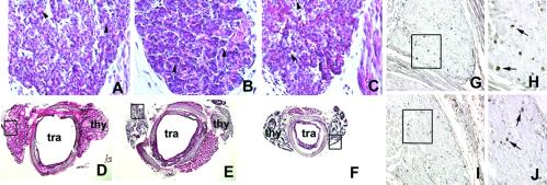
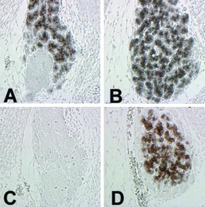
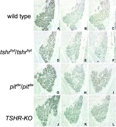
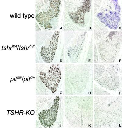
The thyroid-stimulating hormone/thyrotropin (TSH) is the most relevant hormone in the control of thyroid gland physiology in adulthood. TSH effects on the thyroid gland are mediated by the interaction with a specific TSH receptor (TSHR). We studied the role of TSHTSHR signaling on gland morphogenesis and differentiation in the mouse embryo using mouse lines deprived either of TSH (pit(dw)pit(dw)) or of a functional TSHR (tshr(hyt)tshr(hyt) and TSHR-knockout lines). The results reported here show that in the absence of either TSH or a functional TSHR, the thyroid gland develops to a normal size, whereas the expression of thyroperoxidase and the sodium/iodide symporter are reduced greatly. Conversely, no relevant changes are detected in the amounts of thyroglobulin and the thyroid-enriched transcription factors TTF-1, TTF-2, and Pax8. These data suggest that the major role of the TSH/TSHR pathway is in controlling genes involved in iodide metabolism such as sodium/iodide symporter and thyroperoxidase. Furthermore, our data indicate that in embryonic life TSH does not play an equivalent role in controlling gland growth as in the adult thyroid.


Figures

Fig 1.
Fig 1.
Hematoxylin/eosin stain of wild-type and mutant mouse thyroid gland and BrdUrd incorporation in developing mouse thyroid gland. (AC) Sagittal sections of E17 wild-type (A), tshrhyt/tshrhyt (B), and pit1dw/pit1dw (C) mouse embryos. The black arrowheads in AC point to small follicles. These data were representative of at least two independent experiments performed on embryos coming from different littermates. As controls, wild-type C57BL/6 embryos were used. (DF) Transversal sections of thyroid gland and trachea of wild-type (D), tshrhyt/tshrhyt (E), and pit1dw/pit1dw (F) of 2-month-old mice. thy, thyroid; tra, trachea. These data were representative of three independent experiments (two homozygous males and one homozygous female for both tshrhyt and pitdw strains). The mice were generated in different littermates. As controls, wild-type C57BL/6 mice were used. (GJ) Sagittal sections of E16.5 wild-type (G and H) and tshrhyt/tshrhyt (I and J) embryos. H and J show higher magnification of the boxed areas in G and H, respectively. These data are representative of three independent experiments. Embryos from untreated pregnant mice at the same days of gestation were used as a negative control. Cells that incorporated BrdUrd are visible as black dots (arrows).
Fig 2.
Fig 2.
Onset of NIS expression in the developing mouse thyroid gland. Serial sagittal sections of E14.5 (A and C) and E16 (B and D) embryos were stained with anti-Tg (A and B) and anti-NIS (C and D) antibodies. These data are representative of three independent experiments.
Fig 3.
Fig 3.
Expression of thyroid transcription factors in developing mouse thyroid gland. Serial sagittal sections of wild-type (AC), tshrhyt/tshrhyt (DF), pit1dw/pit1dw (GI), and TSHR-KO (JL) E17.5 embryos were stained with anti-TTF-1 (A, D, G, and J), anti-TTF-2 (B, E, H, and K), and anti-Pax8 (C, F, I, and L) antibodies. These data are representative of at least two independent experiments. As controls, wild-type C57BL/6 embryos were used.
Fig 4.
Fig 4.
Expression of Tg, NIS, and TPO in developing mouse thyroid gland. Serial sagittal sections of wild-type (AC), tshrhyt/tshrhyt (DF), pit1dw/pit1dw (GI), and TSHR-KO (JL) E17.5 embryos were stained with anti-Tg (A, D, G, and J) and anti-NIS (B, E, H, and K) or hybridized with TPO antisense probe (C, F, I, and L). These data are representative of at least two independent experiments. As controls, wild-type C57BL/6 embryos were used.
Fig 5.
Fig 5.
Thyroid-specific down-regulation of NIS protein. Sagittal sections of wild-type (A and E), tshrhyt/tshrhyt (B and F), pit1dw/pit1dw (C and G), and TSHR-KO (D and H) E17.5 thyroids and salivary glands were stained with anti-NIS antibody. Salivary glands shown in EH belong to the embryos shown in AD, respectively. NIS expression is very scarce in the thyroid of mutant embryos. In contrast, a clear signal is present in the salivary glands. These data are representative of at least two independent experiments. As controls, wild-type C57BL/6 embryos were used.
Fig 6.
Fig 6.
Structure of the titf1 locus modified by homologous recombination and expression of the targeted allele. (A) The black box indicates titf1 exons, and the black bar indicates the probe used to identify the targeted allele. (B) Human TSHR RNA expression in lung of adult targeted mice. Lanes 1 and 3, lung RNA from titf1+/KI-TSHR* animals; lanes 2 and 4, lung RNA from wild-type animals. (Left) Hybridization with human TSHR probe. (Right) Hybridization with rat TTF-1 probe.
Fig 7.
Fig 7.
Rescue of NIS expression. Serial sagittal sections of adult wild-type (A and D), pit1dw/pit1dw (B and E), and titf1+/KI-TSHR*;pit1dw/pit1dw (C and F) thyroids were stained with anti-NIS antibody (AC) or hematoxylin/eosin (DF). NIS expression is absent in the thyroid gland of pit1dw/pit1dw (B), whereas its expression is rescued in double mutants titf1+/KI−TSHR*;pit1dw/pit1dw (C). These data are representative of at least two independent experiments.
Fig 8.
Fig 8.
Analysis of titf1+/KI-TSHR* E13.5 embryos. Serial sagittal sections were stained with anti-TTF-1 (A) or anti-NIS (B) or hybridized with TPO antisense probe (C). These data are representative of at least two independent experiments.

Similar articles

Cited by

References

    1. Kaufman M. H. & Bard, J., (1999) The Anatomic Basis of Mouse Development (Academic, San Diego), pp. 165–166.
    1. Di Lauro R. & De Felice, M. (2001) in Endocrinology, eds. DeGroot, L. J. & Jameson, J. L. (Saunders, Philadelphia), Vol. 2, pp. 1268–1278.
    1. Lazzaro D., Price, M., De Felice, M. & Di Lauro, R. (1991) Development (Cambridge, U.K.) 113 1093-1104. - PubMed
    1. Zannini M., Avantaggiato, V., Biffali, E., Arnone, M. I., Sato, K., Pischetola, M., Taylor, B. A., Phillips, S. J., Simeone, A. & Di Lauro, R. (1997) EMBO J. 16 3185-3197. - PMC - PubMed
    1. Plachov D., Chowdhury, K., Walther, C., Simon, D., Guenet, J. L. & Gruss, P. (1990) Development (Cambridge, U.K.) 110 643-651. - PubMed

Publication types

MeSH terms

Substances