Comparative Study
. 2002 Nov 26;99(24):15566-71.
doi: 10.1073/pnas.242321099. Epub 2002 Nov 13.

RAG-dependent peripheral T cell receptor diversification in CD8+ T lymphocytes

Affiliations
Comparative Study

RAG-dependent peripheral T cell receptor diversification in CD8+ T lymphocytes

Pau Serra et al. Proc Natl Acad Sci U S A. .

Abstract

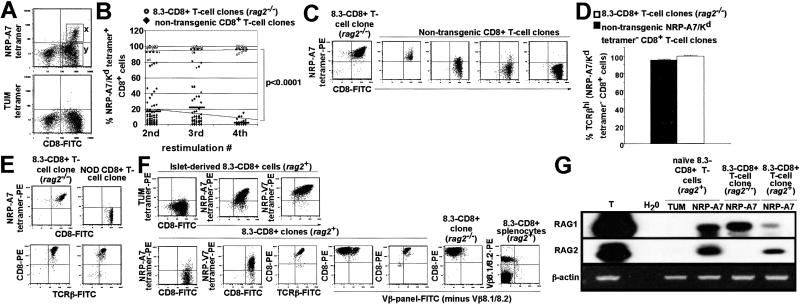
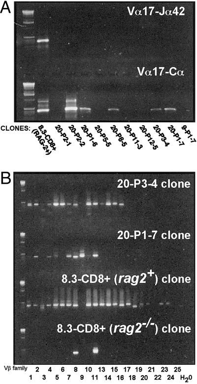
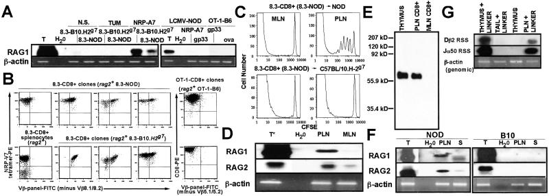
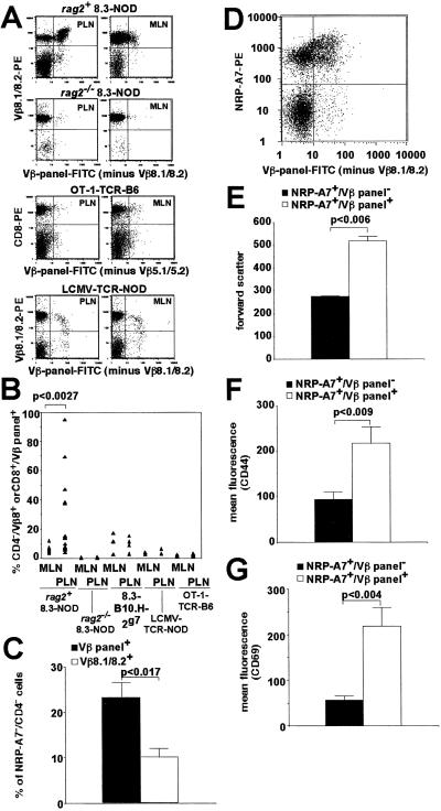
Rearrangement of T cell receptor (TCR) genes is driven by transient expression of V(D)J recombination-activating genes (RAGs) during lymphocyte development. Immunological dogma holds that T cells irreversibly terminate RAG expression before exiting the thymus, and that all of the progeny arising from mature T cells express the parental TCRs. When single pancreatic islet-derived, NRP-A7 peptide-reactive CD8(+) T cells from nonobese diabetic (NOD) mice were repeatedly stimulated with peptide-pulsed dendritic cells, daughter T cells reexpressed RAGs, lost their ability to bind to NRP-A7K(d) tetramers, ceased to transcribe tetramer-specific TCR genes, and, instead, expressed a vast array of other TCR rearrangements. Pancreatic lymph node (PLN) CD8(+) T cells from animals expressing a transgenic NRP-A7-reactive TCR transcribed and translated RAGs in vivo and displayed endogenous TCRs on their surface. RAG reexpression also occurred in the PLN CD8(+) T cells of wild-type NOD mice and could be induced in the peripheral CD8(+) T cells of nondiabetes-prone TCR-transgenic B10.H2(g7) mice by stimulation with peptide-pulsed dendritic cells. In contrast, reexpression of RAGs could not be induced in the CD8(+) T cells of B6 mice expressing an ovalbumin-specific, K(b)-restricted TCR, or in the CD8(+) T cells of NOD mice expressing a lymphocytic choriomeningitis virus-specific, D(b)-restricted TCR. Extra-thymic reexpression of the V(D)J recombination machinery in certain CD8(+) T cell subpopulations, therefore, enables further diversification of the peripheral T cell repertoire.


Figures

Fig 1.
Fig 1.
Loss of tetramer reactivity by NRP-A7-specific CD8+ clones. (A) Pancreatic islets from nondiabetic NOD mice (n = 16–20 mice per experiment; two experiments) were cultured in the presence of rIL-2 for 7 days, and the islet-derived NRP-A7 tetramer-positive (x) and tetramer-negative (y) CD8+ cells were sorted by FACS at one cell per well. (B) Summary of NRP-A7 tetramer reactivity of T cell clones derived from single, originally tetramer-positive cells. Clones were derived from NOD (n = 77, 46, and 21 clones at each restimulation) or rag2−/− 8.3-NOD islets (n = 15–18 clones per restimulation) upon stimulation with NRP-A7-pulsed DCs. (C) Representative tetramer-staining patterns for clones derived from tetramer-positive cells. (D) TCR levels on clones from NOD or rag2−/− 8.3-NOD mice (n = 10 and 8, respectively). (E) Representative tetramer/TCRβ-staining patterns. (F) rag2+ 8.3-CD8+ clones express endogenous TCRs and decrease their avidity for NRP-A7 upon stimulation with NRP-A7-pulsed DCs. (Upper) Images correspond to an islet-derived 8.3-CD8+ T cell line before stimulation in vitro. (Lower) Images correspond to 8.3-CD8+ clones grown on NRP-A7-pulsed DCs or to splenic 8.3-CD8+ T cells. Dead cells were gated out. (G) Expression of RAG mRNAs by 8.3-CD8+ MLN cells and rag2+ and rag2−/− 8.3-CD8+ clones (harvested 1–2 weeks after the first or second restimulations, respectively) within 3 h of stimulation with NRP-A7-pulsed DCs. T, thymus RNA control.
Fig 2.
Fig 2.
TCR repertoire of clones arising from tetramer-positive precursors, as analyzed by RT-PCR. (A) NRP-A7 tetramer-negative clones do not transcribe Vα17-Jα42 mRNA (Upper), but a subset does transcribe at least some Vα17+ TCRα rearrangements (Lower). (B) NRP-A7 tetramer-negative clones express multiple Vβ elements. TCRβ cDNAs of clones were amplified with Vβ- and Cβ-specific primers. The Vβ11-binding primer crossreacted with Vβ8 in this experiment.
Fig 3.
Fig 3.
Expression of RAGs by NRP-A7-reactive CD8+ T cells in vitro and in vivo. (A) Expression of RAG1 mRNA by 8.3-, OT-1-, and LCMV-TCR-transgenic CD8+ T cells upon in vitro stimulation with NRP-A7, ova, or gp33-peptide-pulsed DCs (3 h), respectively. Negative control peptides: TUM, gp33, and NRP-A7, respectively. (B) 8.3-CD8+ clones from 8.3-B10.H2g7 mice express endogenous TCRs upon repeated stimulation with NRP-A7-pulsed DCs. Panels correspond to splenic 8.3-CD8+ cells from a rag2−/− 8.3-NOD mouse (control), a Vβ panel clone from the spleen of a rag2+ 8.3-NOD mouse, and Vβ panel+ clones from the spleens of rag2+ 8.3-NOD or 8.3-B10.H2g7 mice (experiments independent of those shown in Fig. 1). (C) Proliferation of naïve, CFSE-labeled 8.3-CD8+ T cells in the PLNs of NOD but not of B10.H-2g7 mice. (D) Transcription of RAGs by PLN 8.3-CD8+ T cells. CD8+ cells were purified from the PLNs or MLNs of 3–10 8.3-NOD mice per experiment. Thymus (T) RNA was prepared from crude thymus lysates. RT-PCR products [5% for the thymus RAG2 RT-PCR shown (T*) and 15% for all other RT-PCR products] were probed with RAG1 and RAG2 cDNAs. (E) Immunoprecipitation Western blot for RAG2. RAG2 was immunoprecipitated from purified 8.3-CD8+ T cells or whole thymus (250 μg of protein). (F) RAG transcription in PLN and splenic (S) T cells from NOD mice (3–10 mice per experiment). (G) RSS-specific DNA breaks at Dβ and Jα loci in PLN 8.3-CD8+ cells.
Fig 4.
Fig 4.
Expression of secondary TCR rearrangements by 8.3-CD8+ T cells in vivo. (A and B) RAG-dependent expression of secondary TCRβ chains on PLN 8.3-CD8+ cells. Data correspond to PLN and MLN cells from 8.3-NOD (n = 17 and 15, respectively), rag2−/− 8.3-NOD (n = 5 and 6), 8.3-B10.H2g7 (n = 5 and 4), OT-1-B6 (n = 3), and LCMV-NOD mice (n = 3; ≥10 weeks of age). Each triangle corresponds to a different sample. (C) Average percentage of tetramer-negative cells within CD4/Vβ panel+ and CD4/Vβ8.1/8.2+ PLN cells of 8.3-NOD mice (n = 5 mice). (D) Representative tetramer reactivity of CD4/Vβ panel+ and CD4/Vβ panel PLN cells. (E–G) Tetramer-positive/Vβ panel+ cells are larger (E) and express higher levels of CD44 (F) and CD69 (G) than tetramer-positive/Vβ panel cells. Data correspond to eight (E and F) and seven (G) 8.3-NOD mice per group.

Similar articles

Cited by

References

    1. Fugmann S., Lee, A., Shokett, P., Villey, I. & Schatz, D. (2000) Annu. Rev. Immunol. 18 495-527. - PubMed
    1. Nagaoka H., Yu, W. & Nussenzweig, M. (2000) Curr. Opin. Immunol. 12 187-190. - PubMed
    1. Turka L., Schatz, D., Oettinger, M., Chun, J., Gorka, C., Lee, K., McCormack, W. & Thompson, C. (1991) Science 16 778-781. - PubMed
    1. Borgulya P., Kishi, H., Uematsu, Y. & Boehmer, H. v. (1992) Cell 69 529-537. - PubMed
    1. Monroe R., Seidl, K., Gaertner, F., Han, S., Chen, F., Sekiguchi, J., Wang, J., Ferrini, R., Davidson, L., Kelsoe, G. & Alt, F. (1999) Immunity 11 201-212. - PubMed

Publication types

MeSH terms